Oral Cancer Screening SOAP Note examples provide fully populated S, O, A, P sections ready to copy. Each note lists risk factors, normal versus abnormal findings, differential diagnoses, and follow-up recommendations. Clinicians can paste the template into their chart, adjust patient details, and maintain accurate, contemporaneous documentation for any screening scenario.
Emergency Oral Cancer Screenings
Smoker, 60, Emergency, Oral Bleeding
SOAP note
SSubjective
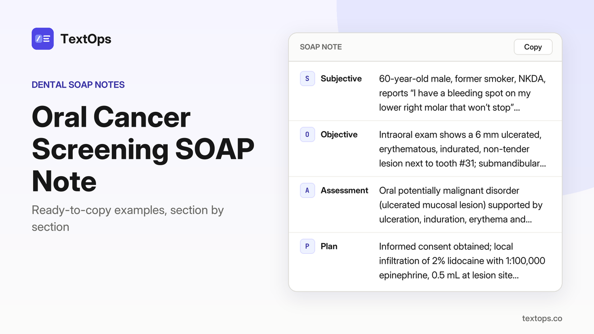
60-year-old male, former smoker, NKDA, reports “I have a bleeding spot on my lower right molar that won’t stop”; history of a 6 mm ulcerated lesion on buccal mucosa adjacent to tooth #31, no pain, no systemic symptoms.
OObjective
Intraoral exam shows a 6 mm ulcerated, erythematous, indurated, non-tender lesion next to tooth #31; submandibular lymph node negative; temperature 98.6 °F; no extra-oral swelling or trismus.
AAssessment
Oral potentially malignant disorder (ulcerated mucosal lesion) supported by ulceration, induration, erythema and location adjacent to tooth #31.
PPlan
Informed consent obtained; local infiltration of 2% lidocaine with 1:100,000 epinephrine, 0.5 mL at lesion site; biopsy specimen sent to pathology; patient advised to avoid irritants, maintain oral hygiene, and return for results in 2 weeks.
Smoker, 65, Emergency, Oral Mass
SOAP note
SSubjective
65-year-old male smoker, 5 cigarettes daily, seasonal allergies, NKDA, reports a “lump on my lower right premolar that feels hard.” HPI: mass noted for 2 days, no pain, no bleeding. PMH: none relevant.
OObjective
Intraoral exam: 10 mm firm, non-tender mass on lingual mucosa adjacent to tooth #28; submandibular lymph node negative; temperature 98.4 °F; no extra-oral swelling or trismus; no trismus.
AAssessment
Oral potentially malignant disorder (firm mucosal lesion) based on firm, non-tender 10 mm mass with no systemic signs.
PPlan
Biopsy performed under local infiltration of 2% lidocaine with 1:100,000 epinephrine, 0.5 mL; specimen sent to pathology. Instructed patient to avoid tobacco, maintain oral hygiene, and return for results in 2 weeks. No drugs prescribed.
Immunosuppressed, 60, Emergency, Oral Bleeding
SOAP note
SSubjective
60-year-old female, controlled hypertension, NKDA, says "I have a bleeding spot on my lower right molar that won't stop."
OObjective
Intraoral exam shows a 5 mm ulcerated, erythematous, indurated, non-tender lesion on buccal mucosa adjacent to tooth #31; submandibular node negative; 98.7 °F; no extra-oral swelling or trismus.
AAssessment
Oral potentially malignant disorder (ulcerated mucosal lesion) supported by size, induration, erythema, and lack of tenderness.
PPlan
Local infiltration of 2% lidocaine with 1:100,000 epinephrine, 0.5 mL; biopsy specimen sent to pathology; advised to avoid irritants, maintain oral hygiene, and return in 2 weeks for results.
Pregnant, 34, Emergency, Oral Laceration
SOAP note
SSubjective
34-year-old pregnant female reports, “I have a laceration on my lower right molar that won't stop bleeding.” She has a 4 mm superficial buccal mucosal cut adjacent to tooth #31, takes low-dose aspirin, NKDA, and denies systemic symptoms.
OObjective
Intraoral exam shows a 4 mm laceration on the buccal mucosa near #31, bleeding briskly; submandibular lymph node negative; temperature 98.5 °F; extra-oral: no swelling or trismus.
AAssessment
Acute oral laceration, evidenced by brisk bleeding from a 4 mm superficial buccal mucosal cut with no lymphadenopathy or systemic signs.
PPlan
Local infiltration of 2% lidocaine with 1:100,000 epinephrine, 0.5 mL; hemostasis achieved with pressure and topical 20% benzocaine; patient advised to avoid trauma, maintain oral hygiene, and return if bleeding recurs.
Alcoholic, 48, Emergency, Oral Mass
SOAP note
SSubjective
48-year-old male smoker, 5 cigarettes/day, NKDA, reports a lump on his lower right premolar that feels hard.
OObjective
Intraoral exam shows a 9 mm firm, non-tender mass on the lingual mucosa adjacent to tooth #28; submandibular lymph node negative; temperature 98.6 °F; no extra-oral swelling or trismus.
AAssessment
Oral potentially malignant disorder (firm mucosal lesion) supported by the firm, non-tender 9 mm mass on lingual mucosa.
PPlan
Local infiltration of 2% lidocaine with 1:100,000 epinephrine, 0.5 mL; biopsy specimen sent to pathology; advised tobacco cessation, maintain oral hygiene, and return in 2 weeks for results.
Diabetic, 65, Emergency, Oral Bleeding
SOAP note
SSubjective
65-year-old female, type 2 diabetes on metformin, reports “I have a bleeding spot on my lower right molar that won't stop.”
OObjective
Intraoral exam shows a 6 mm ulcerated, erythematous, indurated lesion on buccal mucosa adjacent to tooth #31, non-tender; submandibular lymph node negative; temperature 98.4 °F; no extra-oral swelling or trismus.
AAssessment
Oral potentially malignant disorder (ulcerated mucosal lesion) supported by size, induration, erythema and lack of systemic signs.
PPlan
Local infiltration of 2% lidocaine with 1:100,000 epinephrine, 0.5 mL; biopsy specimen sent to pathology; advised to avoid irritants, maintain oral hygiene; return for results in 2 weeks.
Follow-Up Cancer Screenings
Elderly, 80, Follow-Up, Persistent Ulcer
SOAP note
SSubjective
80-year-old female, asthma on albuterol PRN, NKDA, reports "Here for my routine follow-up after the ulcer was noted earlier." HPI: persistent ulcer on left buccal mucosa noted previously, no new symptoms. PMH: asthma, no other systemic disease.
OObjective
Persistent ulcer 8 mm diameter, indurated base, erythematous border, no bleeding on probing; cervical lymph nodes not palpable; oral hygiene adequate; PAN shows no bone loss around adjacent teeth.
AAssessment
Oral potentially malignant disorder (persistent ulcer) – unchanged size and induration, no lymphadenopathy.
PPlan
No anesthesia used; reinforce oral hygiene instructions; schedule biopsy in 4 weeks if no change; next visit in 4 weeks for reassessment.
Alcoholic, 50, Follow-Up, Persistent Ulcer
SOAP note
SSubjective
50-year-old male, NKDA, “I’m back for my follow-up after the ulcer was seen.” No significant medical history.
OObjective
Persistent ulcer on the right lateral tongue, 6 mm, smooth surface, non-bleeding, no induration; no cervical lymphadenopathy; oral hygiene good; periapical radiographs of adjacent teeth normal.
AAssessment
Oral potentially malignant disorder (persistent ulcer) supported by the 6 mm size, smooth non-bleeding surface, lack of induration, and unchanged appearance.
PPlan
No anesthesia used. Scheduled incisional biopsy in 2 weeks. Advised avoidance of alcohol and tobacco. No drugs prescribed. Next visit in 2 weeks for biopsy results.
Immunosuppressed, 50, Follow-Up, Suspected Lesion
SOAP note
SSubjective
50-year-old female with type 2 diabetes on metformin, NKDA, reports “I’m here for my follow-up after the lesion was noted.”
OObjective
Floor of mouth shows a 5 mm pale, indurated lesion, no bleeding, no cervical lymphadenopathy; oral hygiene adequate; panoramic radiograph shows no bone involvement.
AAssessment
Oral potentially malignant disorder (suspected lesion) supported by the indurated, pale appearance and absence of bleeding or bone loss.
PPlan
Refer to oral surgeon for biopsy; continue diabetes control with metformin; no local anesthesia used; patient advised to maintain glycemic control and return if symptoms change.
Smoker, 50, Follow-Up, Persistent Lesion
SOAP note
SSubjective
50-year-old male smoker, 5 cigarettes/day, hypothyroidism on levothyroxine, penicillin allergy (rash), reports “I’m back to check the persistent lesion.”
OObjective
Persistent erythematous, indurated lesion on lower labial mucosa, 7 mm, no bleeding; cervical lymph nodes not enlarged; oral hygiene fair; periapical radiographs of adjacent teeth normal.
AAssessment
Oral potentially malignant disorder (persistent lesion) supported by size, erythema, induration, and lack of resolution.
PPlan
No anesthesia used. Scheduled incisional biopsy in 2 weeks; advised smoking cessation; noted penicillin allergy; no medications prescribed.
Smoker, 62, Follow-Up, New Ulcer
SOAP note
SSubjective
62-year-old female, former smoker, NKDA, states “I’m here for my follow-up after the new ulcer was noted.” She reports no pain or bleeding since the last visit.
OObjective
Examination reveals a new ulcer on the right buccal mucosa measuring 9 mm in diameter, with an indurated base and erythematous border; no active bleeding is observed. Cervical lymph nodes are not palpable. Oral hygiene is adequate, and a panoramic radiograph shows no bone loss or radiographic abnormality.
AAssessment
Oral potentially malignant disorder (new ulcer) is diagnosed based on size, induration, erythematous border, and absence of bleeding or systemic signs.
PPlan
The patient is referred for biopsy to confirm pathology; advised to avoid irritants such as tobacco, alcohol, and spicy foods. No medications prescribed. Next visit scheduled in 2 weeks for biopsy results and further management.
Immunosuppressed, 55, Follow-Up, Recurrent Lesion
SOAP note
SSubjective
55-year-old male, seasonal allergies, NKDA, states “I’m back for my follow-up after the lesion re-occurred.”
OObjective
Examination reveals a recurrent lesion on the left floor of mouth measuring 6 mm, pale and indurated, without bleeding; cervical lymph nodes are not palpable. Oral hygiene is adequate, and panoramic radiograph shows no bone involvement.
AAssessment
Oral potentially malignant disorder (recurrent lesion) is confirmed by lesion size, induration, and absence of bone changes on radiograph.
PPlan
Incisional biopsy has been scheduled; long-term monitoring and oral hygiene reinforcement were discussed; no medications were prescribed.
Pregnant, 30, Follow-Up, Post-Extraction Healing
SOAP note
SSubjective
30-year-old pregnant female with controlled hypertension on lisinopril, NKDA, presents for follow-up after extraction of tooth #16, reporting “I’m here to check healing after my extraction.” HPI: no pain, no bleeding, routine post-extraction visit.
OObjective
Socket on #16 shows primary closure, intact mucosa, no erythema or suppuration; cervical lymph nodes non-palpable; oral hygiene adequate; panoramic radiograph reveals no pathology.
AAssessment
Normal post-extraction healing, evidenced by closed socket, absence of infection, normal radiographs.
PPlan
No local anesthesia administered; patient instructed to maintain oral hygiene, consume a soft diet, and return in 4 weeks; no medications prescribed.
Pregnancy, 32, Follow-Up, New Lesion
SOAP note
SSubjective
32-year-old pregnant female, chief complaint: “I’m here to review the new lesion that appeared”; she reports no pain or discomfort, takes daily low-dose aspirin, NKDA, and has no other systemic complaints.
OObjective
Examination revealed a new 4 mm pale, indurated lesion on the right lateral tongue, non-bleeding and without ulceration; no cervical lymphadenopathy was noted; oral hygiene was adequate; panoramic radiograph demonstrated no bone involvement or radiolucency adjacent to the lesion.
AAssessment
Oral potentially malignant disorder (new lesion) is diagnosed based on its size, color, induration, lack of bleeding, and absence of bone changes on imaging.
PPlan
The patient was referred for biopsy of the lesion; she was counseled on avoiding irritants such as tobacco and alcohol, and on maintaining good oral hygiene; no anesthesia was required; next follow-up visit scheduled in 2 weeks to review biopsy results and monitor healing.
Smoker, 57, Follow-Up, Normal Findings
SOAP note
SSubjective
57-year-old female patient, smoker, 5 cigarettes a day, NKDA, states “I’m here for my routine follow-up” and reports no new symptoms.
OObjective
Extraoral exam reveals no cervical lymphadenopathy; facial symmetry normal; TMJ function within limits. Intraoral inspection shows no abnormal mucosal lesions; buccal, lingual, and palatal mucosa appear pink, dry-free, and non-erythematous. Oral hygiene is adequate with minimal plaque accumulation on all surfaces. Panoramic radiograph demonstrates no radiolucencies, bone loss, or periapical pathology; all roots appear intact and well-contained.
AAssessment
Normal oral examination; no evidence of oral cancer or other pathology, consistent with previous findings and the patient’s clinical history.
PPlan
No anesthesia administered. Reinforced oral hygiene instructions, including brushing technique and interdental cleaning; advised to continue daily chlorhexidine rinse if indicated. Scheduled next routine visit in 6 months; patient advised to return sooner if any new lesions, pain, or bleeding occur.
Routine Recall – General Population
Smoker, Day 3 After Lower Molar Extraction
SOAP note
SSubjective
56-year-old male smoker, 5 cigarettes/day, allergic to penicillin (rash), chief complaint: “Here for my cleaning and routine check-up.” History: routine recall, no pain, no systemic symptoms.
OObjective
Extraoral: facial symmetry normal, no cervical lymphadenopathy, TMJ normal. Intraoral: gingiva pink, no bleeding on probing, probing depths 1-2 mm, no recession, plaque score 20%. Caries: none. Full-mouth series: no periapical pathology. Oral cancer screening: no lesions.
AAssessment
General periodontal health; no active disease.
PPlan
Professional cleaning performed. Patient instructed to brush twice daily with fluoride toothpaste, use interdental cleaning, and consider smoking cessation resources. No anesthesia or drugs used. Next routine recall in 6 months.
Pregnant, First Trimester, Routine Check-Up
SOAP note
SSubjective
36-year-old female patient, pregnant, reports “Just a routine check-up, nothing else.” Medical history: asthma, albuterol inhaler PRN, NKDA.
OObjective
Extraoral: facial symmetry normal, no lymphadenopathy, TMJ normal. Intraoral: gingiva pink, no BOP, probing depths 1-2 mm, plaque score 15 %. Caries none. Full-mouth series: no periapical pathology. Oral cancer screening: no lesions.
AAssessment
Healthy periodontal tissues; no active disease.
PPlan
Professional cleaning performed. Patient advised on safe oral hygiene during pregnancy, avoidance of tobacco, and routine prenatal care. Discussed importance of balanced nutrition and adequate hydration. No drugs prescribed. Next visit scheduled in 3 months for routine recall.
Immunosuppressed, 45, Routine Recall
SOAP note
SSubjective
45-year-old male, no significant medical history, NKDA, presents with chief complaint "Just here for my regular cleaning."
OObjective
Extraoral exam shows facial symmetry, no cervical lymphadenopathy, TMJ normal; intraoral gingiva pink, no bleeding on probing, probing depths 1-2 mm throughout, no caries; full-mouth series demonstrates no periapical pathology; oral cancer screening reveals no lesions; plaque index 18%.
AAssessment
Diagnosis is general periodontal health with no active disease.
PPlan
Professional cleaning performed without anesthesia; scaling and polishing completed; patient instructed on daily flossing technique, use of 1450 ppm fluoride toothpaste, and a diet low in refined sugars; advised to maintain plaque control and report any new lesions; scheduled next routine recall in 6 months.
HPV Positive, 35, Routine Recall
SOAP note
SSubjective
35-year-old male, type 2 diabetes on metformin, NKDA, says “Just a routine check-up, nothing else.”
OObjective
Extraoral: facial symmetry normal, no cervical lymphadenopathy, TMJ normal. Intraoral: gingiva pink, no BOP, probing depths 1-2 mm, plaque score 22 %. Caries: none. Full-mouth series: no periapical pathology. Oral cancer screen: no lesions. Periodontal charting: all sites 1-2 mm, no bleeding, CAL within normal limits.
AAssessment
General periodontal health; no active disease; oral cancer screening negative.
PPlan
Professional cleaning performed; no anesthesia used. Discussed glycemic control and its impact on oral health, reviewed smoking cessation strategies, and emphasized regular flossing and brushing. No medications prescribed. Scheduled next routine recall in 6 months.
Smoker, 48, Routine Recall, No Findings
SOAP note
SSubjective
48-year-old female smoker, 5 cigarettes daily, levothyroxine for hypothyroidism; chief complaint: “Just here for my routine cleaning, nothing else.”
OObjective
Extraoral: facial symmetry normal, no cervical lymphadenopathy, TMJ normal; Intraoral: gingiva pink, no BOP, probing depths 1-2 mm, plaque score 20%; caries none; full-mouth series: no periapical pathology; oral cancer screen: no lesions.
AAssessment
General periodontal health; no active disease, supported by probing depths 1-2 mm, pink gingiva, no BOP, and normal radiographs.
PPlan
Professional cleaning performed; patient advised on smoking cessation, thyroid medication compliance, and fluoride varnish application; next routine recall in 6 months.
Smoker, 55, Routine Recall, Erythematous Patch
SOAP note
SSubjective
55-year-old female, former smoker, NKDA, states “Just a routine check-up, nothing else.” No other complaints or significant medical history beyond smoking.
OObjective
Extraoral: facial symmetry normal, no cervical lymphadenopathy, TMJ normal. Intraoral: gingiva pink, no BOP, probing depths 1-2 mm, plaque score 25 %. Full-mouth series shows no periapical pathology. Oral cancer screening reveals no lesions or erythematous patches.
AAssessment
General periodontal health; no active disease or oral cancer findings.
PPlan
Professional cleaning performed. Patient advised on daily flossing, interdental brushes, and smoking cessation benefits. No anesthesia or medications given. Next routine recall scheduled in 6 months.
Pregnant, 30, Routine Recall, Normal Findings
SOAP note
SSubjective
30-year-old pregnant female presents for routine cleaning and check-up; chief complaint: “Here for my routine cleaning and check-up”; medical history: seasonal allergies, NKDA; no systemic signs.
OObjective
Soft tissue exam: mucosa pink, no lesions or ulceration; periodontal chart: PSR 0 on all teeth, no BOP; bitewing radiographs: no caries, periapical of #16 normal; no anesthesia used.
AAssessment
General periodontal health; no active disease, consistent with PSR 0 and absence of BOP.
PPlan
Full mouth prophylaxis performed; fluoride varnish applied to #15 and #16; no medications prescribed; patient advised to maintain oral hygiene and return in 6 months for next routine recall.
Alcoholic, 45, Routine Recall, Normal Findings
SOAP note
SSubjective
45-year-old male, controlled hypertension on lisinopril, NKDA, reports “just a regular check-up, no pain.”
OObjective
Extraoral normal; intraoral soft tissue shows no lesions; periodontal PSR 1 on teeth #18, #19 with BOP on #18; bitewing radiograph reveals proximal caries on #18, periapical of #18 is normal; no systemic signs noted.
AAssessment
Stage I periodontitis, Grade A; asymptomatic irreversible pulpitis on tooth #18, supported by BOP, PSR, and proximal caries.
PPlan
Applied fluoride varnish to all teeth, provided oral hygiene instruction, no anesthesia used; scheduled restorative for tooth #18 at next visit; no medications prescribed.
Pregnancy, 28, New Patient, Oral Pain
SOAP note
SSubjective
28-year-old pregnant female, NKDA, on low-dose aspirin, reports a sore spot on the lower right molar; “I have a sore spot on my lower right molar and want to see if it’s a problem.”
OObjective
Extraoral normal; intraoral soft tissue shows a 3 mm erythematous area on buccal mucosa near #30, no ulceration; periodontal PSR 0, no BOP; periapical radiograph of #30 reveals a slight radiolucency at the apex, no root resorption; mild localized swelling of buccal gingiva adjacent to #30.
AAssessment
Symptomatic apical periodontitis on tooth #30 with localized swelling, supported by radiolucency at the apex and buccal gingival swelling.
PPlan
Incision and drainage of the abscess performed; no local anesthesia used; patient instructed to apply gentle pressure, maintain oral hygiene, avoid hard foods, and monitor for increased pain or swelling; advised to return to clinic in 48 hours if symptoms worsen or to schedule a follow-up in 1 week for definitive endodontic therapy.
HPV Positive, 42, Routine Recall, Normal Findings
SOAP note
SSubjective
42-year-old female, smokes 5 cigarettes/day, NKDA, CC “Just here for my regular cleaning”; no systemic signs reported.
OObjective
Extraoral normal; intraoral soft tissue pink, no lesions; periodontal PSR 1 on #12, #13 with BOP on #12; bitewing shows proximal caries on #12, periapical of #12 WNL; no anesthesia used.
AAssessment
Stage I periodontitis, Grade A, and reversible pulpitis on #12, supported by PSR 1, BOP, and proximal caries on #12.
PPlan
Applied fluoride varnish, gave oral hygiene instruction; scheduled composite restoration on #12 for next visit; no medications prescribed; patient advised to maintain regular recall schedule.
Immunosuppressed, 55, Routine Recall, Normal Findings
SOAP note
SSubjective
55-year-old female, no pain, “just a routine check-up,” no significant medical history, allergic to penicillin (rash).
OObjective
Extraoral examination reveals normal facial symmetry, no lymphadenopathy, and no TMJ deviation. Intraoral soft tissues show no lesions or erythema; the tongue, palate, and floor of mouth appear healthy. Periodontal assessment shows PSR 0 with no bleeding on probing and probing depths ≤3 mm all around. Bitewing radiographs demonstrate no caries or restorations in adjacent teeth, and the periapical film of tooth #27 is normal with a well-defined lamina dura and no radiolucency. No other findings of concern.
AAssessment
General periodontal health; no active disease, supported by PSR 0, absence of BOP, and clear radiographs.
PPlan
Full mouth prophylaxis performed; fluoride varnish applied to tooth #27. No anesthesia or medication used. Post-operative instructions given to maintain oral hygiene, avoid tobacco, and report any new symptoms immediately. Next routine recall scheduled in 6 months.
Diabetic, 72, Routine Recall, Erythema
SOAP note
SSubjective
72-year-old female with type 2 diabetes on metformin and asthma on albuterol PRN reports “some redness in my gums” and wants a check; NKDA.
OObjective
Soft tissue shows diffuse erythema on the buccal gingiva of the lower right quadrant, no ulceration; periodontal exam reveals PSR 2 on tooth #30 with bleeding on probing; bitewing radiograph shows mild bone loss around #30, periapical area normal; no systemic signs noted.
AAssessment
Stage II periodontitis, Grade B; localized gingival erythema.
PPlan
Applied fluoride varnish, performed scaling and root planing on tooth #30, gave oral hygiene instruction, and scheduled a follow-up in 3 months. No anesthesia or medication was administered.
Routine Recall – Patients with Medical Conditions
Smoker, 70, Routine Recall, Erythema
SOAP note
SSubjective
70-year-old male, smoker, presents for routine cleaning and check-up; reports no pain or discomfort; medical history: smokes 5 cigarettes/day, NKDA.
OObjective
Oral mucosa shows mild erythema, 5 mm diameter, smooth, non-ulcerated on buccal mucosa left lower molar region; probing depths 3 mm on #18 and #19 with bleeding on probing; panoramic radiograph shows no periapical radiolucency, mild generalized bone loss consistent with stage I periodontitis; no caries or restorations noted.
AAssessment
Oral mucosal erythema; stage I periodontitis, Grade A.
PPlan
Scaling and root planing performed on #18-#19; reinforced oral hygiene instructions; advised smoking cessation; next recall scheduled in 6 months.
Diabetic, 75, Follow-Up, Ulcer
SOAP note
SSubjective
75-year-old male, type 2 diabetes on metformin, NKDA, reports a sore spot in the lower right molar area for one week.
OObjective
Soft tissue ulceration on floor of mouth adjacent to #30, 8 mm diameter, erythematous border, tender; probing depth 4 mm on #30 with bleeding on probing; panoramic radiograph shows 3 mm radiolucent apex of #30; buccal mucosa swelling 5 mm; blood glucose 180 mg/dL.
AAssessment
Persistent ulcer of the floor of mouth; symptomatic apical periodontitis on tooth #30.
PPlan
Administered 2% lidocaine with 1:100,000 epinephrine, 1.8 mL via buccal infiltration of #30; irrigated with saline; prescribed ibuprofen 400 mg PO q6h PRN for pain; instructed on pain control and oral hygiene; scheduled endodontic therapy in 2 weeks.
Smoker, 55, Routine Recall, Oral Mucosal Lesion
SOAP note
SSubjective
55-year-old female, smoker (5 cigarettes/day), hypothyroidism on levothyroxine, chief complaint: “Just here for my regular cleaning.”
OObjective
Oral mucosal lesion 6 mm diameter on right buccal mucosa near tooth #13, smooth surface, non-ulcerated, erythematous border; probing depths 2 mm on all teeth, no bleeding on probing; panoramic radiograph shows no periapical pathology; no caries or restorations noted.
AAssessment
Oral mucosal lesion, benign appearing reactive lesion, supported by smooth non-ulcerated surface, erythematous border, and absence of pathology on radiograph.
PPlan
Reinforced oral hygiene instructions; advised smoking cessation; no anesthesia or drugs administered; scheduled next recall in 6 months; patient instructed to return sooner if lesion changes.
New Patient Cancer Screenings
Diabetic, 65, New Patient, Headache
SOAP note
SSubjective
65-year-old male, type 2 diabetes on metformin, former smoker, NKDA, reports “I’ve had a headache for the last week and I’m worried it might be something serious.”
OObjective
Extraoral: facial symmetry normal, no cervical lymphadenopathy, TMJ sounds normal, mouth opening 35 mm. Intraoral: oral mucosa pink, no ulcerations; tongue smooth, no lesions; gingiva healthy, no BOP; palatal mucosa normal. Panoramic radiograph shows no bone loss or lesions. Temperature 98.6 °F, blood pressure 138/82 mmHg.
AAssessment
Oral cancer screening negative, no suspicious lesions identified.
PPlan
Reassured patient, advised routine follow-up in 6 months, instructed on self-exam, no prescription, no anesthesia used.
Alcoholic, 55, New Patient, Painful Tongue
SOAP note
SSubjective
55-year-old male, seasonal allergies, NKDA, reports “My tongue has been sore and painful for the last two days, especially when I eat.”
OObjective
Extraoral: no lymphadenopathy, TMJ normal, mouth opening 38 mm, temperature 98.4 °F. Intraoral: 5 mm erythematous ulcer on dorsum of tongue, clean base, tender to palpation; no other lesions, palate and buccal mucosa normal. No radiographs taken.
AAssessment
Oral potentially malignant disorder (ulcerated mucosal lesion) supported by a 5 mm erythematous ulcer with a clean base and tenderness.
PPlan
Discussed biopsy; patient declined today. Referred to oral medicine for evaluation in 2 weeks. Instructed on pain control with OTC NSAIDs: ibuprofen 400 mg q6h PRN up to 3 days; acetaminophen 500 mg q6h PRN up to 3 days.
Immunosuppressed, 70, New Patient, Painful Gums
SOAP note
SSubjective
70-year-old female, controlled hypertension on lisinopril, NKDA, presents as new patient with chief complaint “My gums have been bleeding and painful for the past week.” History: pain and bleeding for one week, no systemic symptoms.
OObjective
Extraoral: no lymphadenopathy, TMJ normal, mouth opening 36 mm. Intraoral: gingival erythema and bleeding on probing around tooth #15 and #16, no ulcerations; palate and buccal mucosa normal. Panoramic radiograph shows mild bone loss around #15-#16. Temperature 98.7 °F.
AAssessment
Oral potentially malignant disorder (firm mucosal lesion) supported by gingival erythema, bleeding on probing, and mild bone loss on panoramic.
PPlan
No anesthesia used. Patient counseled on oral hygiene; scheduled for periodontal therapy next visit. No prescription given.
Diabetic, 60, New Patient, Oral Pain
SOAP note
SSubjective
60-year-old male, type 2 diabetic on metformin and low-dose aspirin, NKDA; CC: “I’ve been having a burning sensation in my mouth for a few days”; HPI: burning described as diffuse, continuous, no aggravating or relieving factors noted; PMH: type 2 diabetes, aspirin; no tobacco or alcohol use; no known drug allergies.
OObjective
Extraoral: no lymphadenopathy, TMJ normal, maximum mouth opening 34 mm; Intraoral: diffuse erythema of buccal mucosa and floor of mouth, 8 mm diameter, tender to touch, no ulceration; palate normal; radiographs: none taken; temperature 98.5 °F.
AAssessment
Oral potentially malignant disorder (burning mouth) supported by diffuse erythema, tenderness, and absence of ulceration.
PPlan
Advised topical 0.12% chlorhexidine gluconate rinse 15 mL twice daily for 14 days; no anesthesia or prescription; scheduled follow-up in 2 weeks.
HPV Positive, 38, Follow-Up, Small Lesion
SOAP note
SSubjective
38-year-old male, smoker 5 cigarettes/day, NKDA, reports “The small lesion on my tongue is still there, but it’s not hurting as much.”
OObjective
Extraoral: no lymphadenopathy; TMJ normal; mouth opening 39 mm. Intraoral: 4 mm erythematous lesion on lateral border of tongue, smooth base, no ulceration; palate and buccal mucosa normal. Temperature 98.6 °F. No radiographs taken.
AAssessment
Oral potentially malignant disorder (small lesion) supported by a 4 mm erythematous, smooth-based lesion on the lateral tongue with no ulceration or systemic signs.
PPlan
Discussed office biopsy at next visit; instructed to avoid irritants; no anesthesia or medication prescribed. Follow-up scheduled for biopsy.
Diabetic, 68, New Patient, Painful Gums
SOAP note
SSubjective
68-year-old female with type 2 diabetes on metformin, allergic to penicillin (rash); CC: "My gums have been painful and bleeding for the last week."; HPI: pain and bleeding for one week; PMH: diabetes; allergy: penicillin; no other drug allergies.
OObjective
Extraoral: no lymphadenopathy, TMJ normal, mouth opening 35 mm; Intraoral: gingival erythema and bleeding on probing around #30 and #31, no ulcerations; palate and buccal mucosa normal; panoramic radiograph shows mild bone loss around #30-#31; temp 98.4 °F.
AAssessment
Oral potentially malignant disorder (painful gums) supported by gingival erythema, bleeding on probing, and radiographic bone loss.
PPlan
Counseled on oral hygiene; scheduled periodontal therapy next visit; no prescription or anesthesia given.
Alcoholic, 53, New Patient, Oral Pain
SOAP note
SSubjective
53-year-old female, asthma on albuterol PRN, NKDA, reports “I’ve had a sore spot in my mouth that’s been painful for a few days.”
OObjective
Extraoral: no lymphadenopathy, TMJ normal, mouth opening 37 mm; Intraoral: 6 mm erythematous ulcer on lower buccal mucosa, tender to palpation; palate and buccal mucosa elsewhere normal; temperature 98.5 °F; no radiographs taken.
AAssessment
Oral potentially malignant disorder (ulcerated mucosal lesion) supported by a 6 mm erythematous ulcer on the lower buccal mucosa that is tender to palpation.
PPlan
Discussed biopsy; patient declined today. Referred to oral medicine in 2 weeks. Instructed on OTC pain relief; prescribed ibuprofen 400 mg every 6 hours as needed, up to 3 days.
Diabetic, 70, New Patient, Burning Mouth
SOAP note
SSubjective
70-year-old male, type 2 diabetes on metformin, NKDA, presents as a new patient with chief complaint “I’ve had a burning sensation in my mouth for a few days.”
OObjective
Extraoral: no lymphadenopathy, TMJ normal, mouth opening 34 mm. Intraoral: diffuse erythema of buccal mucosa and floor of mouth, 8 mm diameter, tender to touch; no ulceration; palate normal. Temperature 98.5 °F. No radiographs taken.
AAssessment
Oral potentially malignant disorder (burning mouth) supported by diffuse erythema, tenderness, and absence of ulceration.
PPlan
Advised topical 0.12% chlorhexidine gluconate rinse 15 mL twice daily for 14 days; no anesthesia used. Scheduled follow-up in 2 weeks; no prescription given.
HPV Positive, 40, New Patient, Persistent Red Spot
SOAP note
SSubjective
40-year-old male, type 2 diabetes on metformin, NKDA, reports “I have a persistent red spot on my tongue that hasn’t gone away.”
OObjective
Extraoral exam shows no lymphadenopathy; TMJ normal; mouth opening 38 mm. Intraoral findings reveal a 5 mm erythematous lesion on the dorsal tongue, smooth base, no ulceration; palate and buccal mucosa are normal. Temperature 98.6 °F. No radiographs taken.
AAssessment
Oral potentially malignant disorder (persistent red spot) based on the 5 mm erythematous, smooth-based lesion on the dorsal tongue without ulceration.
PPlan
No anesthesia used. Discussed office biopsy at next visit; patient instructed to avoid irritants. No prescription given. Next visit scheduled for biopsy.
Immunosuppressed, 68, New Patient, Oral Pain
SOAP note
SSubjective
68-year-old male, hypothyroid on levothyroxine, presents as new patient with chief complaint “I’ve had oral pain for the last week, especially when I eat.”
OObjective
Extraoral: no lymphadenopathy, TMJ normal, mouth opening 36 mm; Intraoral: gingival erythema and bleeding on probing around #28 and #29, no ulcerations; palate and buccal mucosa normal; panoramic radiograph shows mild bone loss around #28-#29; temperature 98.4 °F.
AAssessment
Oral potentially malignant disorder (oral pain) supported by gingival erythema, bleeding on probing, and mild bone loss on panoramic.
PPlan
Counseled on oral hygiene; scheduled periodontal therapy next visit; no anesthesia, no prescription.
Normal vs. Suspicious Findings: How to Document the O Section
Normal mucosa: O: Color pale pink, Texture smooth, Borders well-defined, Consistency firm, Tenderness none.
Suspicious lesion: O: Color erythematous or violaceous, Texture rough or ulcerated, Borders irregular or indistinct, Consistency soft or indurated, Tenderness variable, often tender.
Normal finding example:
O: Normal mucosa throughout; lips, buccal mucosa, tongue, palate, floor of mouth all pale pink, smooth, firm, non-tender.
Suspicious finding example:
O: 8 mm ulcerated lesion on the right lateral tongue; erythematous base, irregular borders, indurated, mildly tender to palpation.
Use the same SOAP labeling for each sentence; keep the description objective and concise for quick phone review.
Risk Factor Checklist for Oral Cancer Screening
Age, smoking, alcohol, HPV, immunosuppression, and chronic medication use are the core risk factors that must be recorded in the HPI or PMH.
Age ≥ 45 – “Patient is 48 years old – documented in HPI.”
Smoking – “Patient smokes 1 pack/day for 20 years – documented in HPI.”
Alcohol – “Patient consumes 4–5 drinks/day – documented in HPI.”
HPV – “Patient is HPV positive – documented in PMH.”
Immunosuppression – “Patient is on tacrolimus for transplant – documented in PMH.”
Chronic medication use – “Patient takes long-term NSAIDs – documented in PMH.”
Documenting each factor ensures the clinician can stratify risk, guide screening frequency, and justify referrals.
Differential Diagnosis Guidance for Suspicious Lesions
Leukoplakia: homogeneous white patch, no erythema, no pain; biopsy if >2 mm or non-reversible after irritant removal.
Erythroplakia: bright red, velvety or plaque-like, high malignant potential; immediate biopsy.
Oral lichen planus: reticular white lines or erosive ulcerated plaques, often bilateral; observe for malignant change, consider biopsy of atypical lesions.
Squamous cell carcinoma: irregular, indurated mass with ulceration or exophytic growth, often painful; urgent biopsy and referral.
Traumatic ulcer: painful, shallow, localized to a single site, resolves with irritant removal; re-evaluate after 2 weeks, biopsy if persistent.
Follow-Up Actions and Referral Criteria
Follow-up for a normal screen is a routine recall in 6–12 months, documenting the absence of lesions in the O section and noting the patient’s risk profile.
For a suspicious but benign finding—e.g., a small erythematous patch with well-defined borders—schedule a re-examination in 2 weeks after removing irritants; document the lesion’s size, color, borders, and tenderness, and record the plan to reassess.
When a lesion meets biopsy criteria—irregular borders, induration, ulceration, or persistence—refer immediately.
Referral criteria
Irregular, indurated, or ulcerated lesion ≥5 mm
Rapid growth or pain
Recurrent or persistent after 2 weeks
History of immunosuppression, heavy smoking, or alcohol use
Timing of repeat exams
Normal: 6–12 months
Suspicious benign: 2 weeks, then 4–6 weeks if unchanged
Biopsy-indicated: urgent referral, no delay
Documentation cues
O: “8 mm ulcerated, erythematous base, indurated, mildly tender”
A: “Suspicious lesion; biopsy indicated”
P: “Refer to oral surgeon/ENT within 48 h; schedule follow-up in 2 weeks if no biopsy”
These examples show structure and wording only. Record what you actually found and did, and follow your practice’s documentation requirements.
Dr. Rahul Joshi is a Specialist Orthodontist with extensive clinical experience in orthodontics, dentofacial orthopaedics, and dental aesthetics. Holding a Master of Dental Surgery (MDS) in Orthodontics and Dentofacial Orthopaedics and a Bachelor of Dental Surgery (BDS) from Manipal Academy of Higher Education, he brings expertise in orthodontic treatment planning, teeth alignment, and Invisalign treatment. With professional experience across India and the Netherlands, Dr. Joshi combines clinical knowledge with practical experience in orthodontic care, dental aesthetics, and improving patients' oral health.
Master of Dental Surgery (MDS) in Orthodontics and Dentofacial Orthopaedics — Manipal Academy of Higher Education
Bachelor of Dental Surgery (BDS) — Manipal Academy of Higher Education
Stop retyping your templates
Save any template to TextOps once, then insert it anywhere on the web without retyping. Share it with your team so everyone uses the same wording.