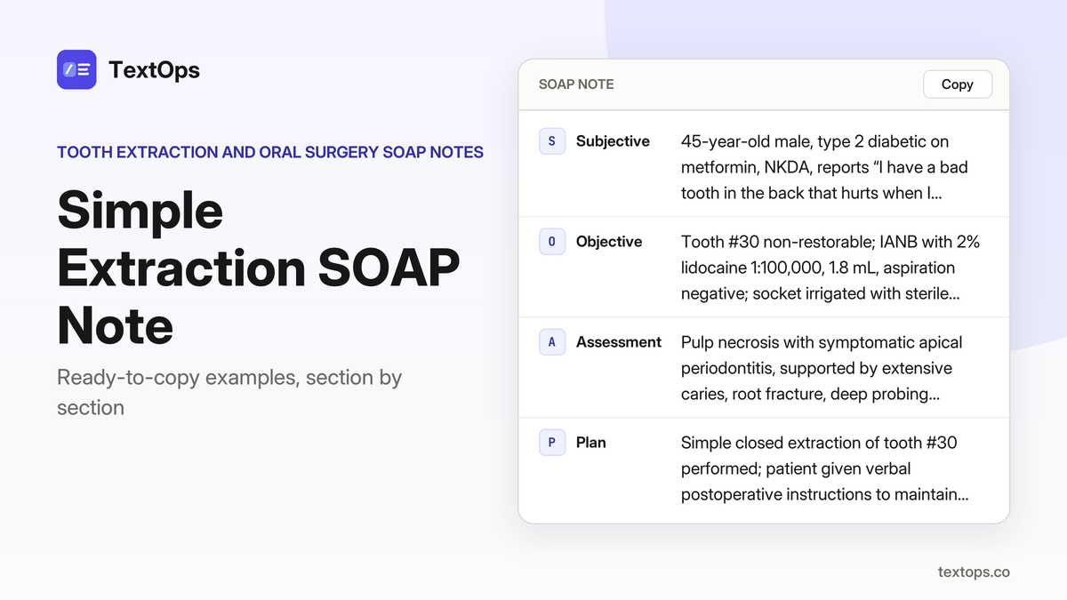
Multiple-tooth extraction SOAP note templates are organized by clinical scenario, from emergency cases to medically complex patients. Each copy-paste block contains complete S, O, A, P sections, ready for immediate use. A mobile-friendly “copy” button lets you grab the template in seconds, even on a handheld device. Scroll down to view the examples and start documenting with precision.
Emergency and Acute Extractions
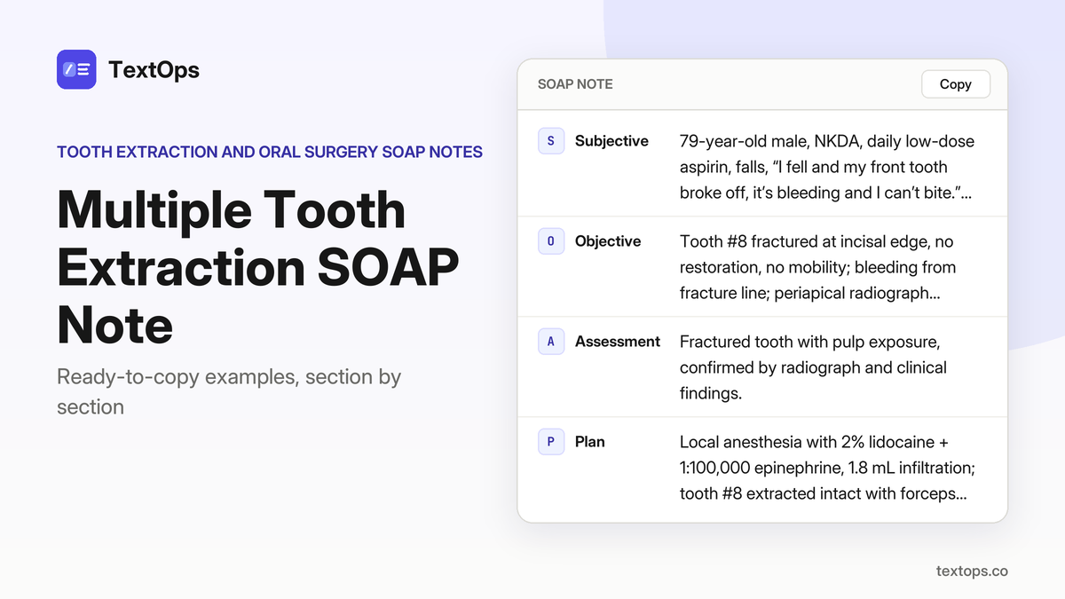
Emergency Extraction of a Fractured Upper Incisor
SOAP note
SSubjective
79-year-old male, NKDA, daily low-dose aspirin, falls, “I fell and my front tooth broke off, it’s bleeding and I can’t bite.” HPI: fracture at incisal edge of tooth #8, bleeding from fracture line, no mobility, intact palatal and labial gingiva, no systemic signs.
OObjective
Tooth #8 fractured at incisal edge, no restoration, no mobility; bleeding from fracture line; periapical radiograph shows fracture line extending into dentin, no periapical radiolucency; vital signs WNL.
AAssessment
Fractured tooth with pulp exposure, confirmed by radiograph and clinical findings.
PPlan
Local anesthesia with 2% lidocaine + 1:100,000 epinephrine, 1.8 mL infiltration; tooth #8 extracted intact with forceps; socket irrigated with saline, gauze pressure applied; verbal POI given: avoid chewing hard foods, apply ice, return if swelling or uncontrolled bleeding. No drugs prescribed.
Extraction of a Tooth with a Large Periapical Abscess
SOAP note
SSubjective
61-year-old male, controlled hypertension on lisinopril, NKDA, reports “my molar is so painful, it’s throbbing and I can’t sleep,” HPI of acute throbbing pain since yesterday, no systemic symptoms.
OObjective
Extraoral: 1 cm buccal mucosa swelling, no lymphadenopathy; intraoral: deep caries to outer dentin, pulpal exposure, mild palatal erythema; radiograph: 4 mm periapical radiolucency at #15 apex, no sinus tract; no swelling beyond buccal mucosa.
AAssessment
Pulp necrosis with acute apical abscess of tooth #15, supported by pulpal exposure, radiolucency, and throbbing pain.
PPlan
Local anesthesia: 2% lidocaine with 1:100,000 epinephrine, 1.8 mL infiltration; extraction of #15 via closed technique, socket irrigated, gauze pressure applied; prescribed ibuprofen 400 mg q6h PRN up to 3 days and acetaminophen 500 mg q6h PRN up to 3 days; POI: soft diet, avoid hot foods, ice packs, keep gauze for 30-60 min, no smoking or rinsing for 24 h; next visit: review healing in 3 days.
Extraction of Third Molar with Jaw Osteomyelitis
SOAP note
SSubjective
38-year-old male, asthma, NKDA, reports “bad swelling in my lower jaw and the tooth hurts a lot.” Fever 38.2 °C, malaise, lymphadenopathy noted.
OObjective
Impacted #30, mesioangular, Pell & Gregory II B; panoramic shows 6 mm radiolucency around apex adjacent to IAC, 2 cm firm palatal swelling, erythematous mucosa, no trismus, temp 38.2 °C.
AAssessment
Impacted third molar with osteomyelitis, supported by radiolucency, swelling, fever, and systemic signs.
PPlan
IANB with 4% articaine 1:100,000, 1.8 mL; flap, bone removal, sectioning, fragment removal, curettage, saline irrigation, gauze + oxidized cellulose hemostasis, 4-0 chromic gut sutures. POI: avoid hard foods, apply ice, take amoxicillin 500 mg q6h PRN for 3 days, ibuprofen 400 mg q6h PRN for 3 days, no smoking. Follow-up in 3 days.
Extraction of Lower Third Molar with Abscess
SOAP note
SSubjective
60-year-old female, former smoker, NKDA, reports “My lower molar is swollen and I can’t eat.” History: deep caries with pulpal exposure on tooth #30, periapical radiograph shows a 5 mm radiolucency, palatal swelling 1.5 cm, tender, erythematous mucosa, no trismus, no fever, no lymphadenopathy.
OObjective
Extraoral: facial symmetry WNL. Intraoral: tooth #30 non-restorable, deep caries, pulpal exposure, palatal swelling 1.5 cm, erythema, tender to palpation. Periodontal: BOP present, probing depths 4–6 mm. Radiograph: 5 mm periapical radiolucency consistent with chronic apical abscess.
AAssessment
Pulp necrosis with chronic apical abscess of tooth #30, supported by deep caries, pulpal exposure, radiolucency, and palatal swelling.
PPlan
Administered 2% lidocaine with 1:100,000 epinephrine, 1.8 mL IANB. Extracted tooth #30 with closed technique, irrigated socket, placed gauze for pressure hemostasis. Prescribed ibuprofen 400 mg q6 h PRN up to 3 days. Provided POI: avoid hard foods, use ice packs, maintain oral hygiene, report bleeding or severe pain. Schedule follow-up in 48 h for healing assessment.
Extraction of Lower Third Molar with Root Fragment
SOAP note
SSubjective
53-year-old female, NKDA, reports “The tooth broke and I can’t bite, there’s a piece stuck in the socket.” She has no medical history or systemic signs; pain is localized to the lower right third molar area.
OObjective
Tooth #30 partially erupted; periapical radiograph shows a 2-mm root fragment remaining in the socket, no periapical radiolucency. Palatal mucosa intact, no swelling, no trismus, no fever, no lymphadenopathy.
AAssessment
Root fragment retained after extraction of tooth #30, supported by radiographic evidence of a fragment in the socket with no surrounding pathology.
PPlan
IANB with 4% articaine + 1:100,000 epinephrine, 1.8 mL, administered. Flap reflected, fragment removed with forceps, socket curetted and irrigated, gauze pressure applied. POI: bite on gauze 30 min, avoid hard foods, maintain soft diet, report swelling or pain. Next visit: follow-up in 7 days.
Special Situations and Complex Cases
First-Time Extraction of 3 Lower Molars
SOAP note
SSubjective
67-year-old male, no significant medical history, allergic to penicillin (rash), chief complaint: “I have a bad tooth that keeps hurting and I can’t eat”; history: deep caries with pulpal exposure on #30, extensive caries with pulpal necrosis and a 4 mm periapical radiolucency on #31, moderate caries without pulpal involvement on #32.
OObjective
Extraoral: normal. Intraoral: #30 deep caries, pulpal exposure, grade 1 mobility; #31 extensive caries, pulpal necrosis, 4 mm periapical radiolucency; #32 moderate caries, no pulpal involvement. Periodontal: BOP absent, PD ≤ 4 mm. Radiographic: PAN shows no root resorption, no sinus communication.
AAssessment
Diagnosis: irreversible pulpitis with pulpal exposure on #30, pulp necrosis with chronic apical periodontitis on #31, caries with no pulpal involvement on #32.
PPlan
Closed-suction extraction of #30, #31, #32 with 2% lidocaine 1:100,000, 1.8 mL IANB; sockets curetted, irrigated with saline; gauze pressure 30 min; POI: bite on gauze, no spitting, soft diet, NSAID as needed, call if pain > moderate or swelling; next visit in 7 days for healing assessment.
Re-evaluation After Failed Root Canal and Extraction of a Mandibular Second Molar
SOAP note
SSubjective
82-year-old male, hypothyroidism on levothyroxine, presents with “The tooth still hurts after the root canal.” History: failed RCT on tooth #18, now symptomatic apical periodontitis with 6 mm periapical radiolucency and middle-third root fracture; swelling 1 cm in the lower left molar region, no trismus, no systemic signs.
OObjective
Extraoral: mild swelling 1 cm in lower left molar area, no facial asymmetry. Intraoral: tooth #18 previously root-canaled, tender on percussion, no mobility. Periodontal: BOP present, probing depths 3–4 mm. Radiographic: PAN shows 6 mm radiolucency at #18, no proximity to inferior alveolar canal. Anesthesia: 2% lidocaine with 1:100,000 epinephrine, 1.8 mL, IANB, aspiration negative.
AAssessment
Symptomatic apical periodontitis of tooth #18, confirmed by clinical tenderness, radiolucency, and root fracture.
PPlan
Closed-suction extraction of #18 performed under IANB; socket irrigated, gauze pressure applied. No medications prescribed. Patient instructed to avoid hard foods for 48 h, maintain oral hygiene, report severe pain or swelling. Next visit: 7-day follow-up to assess healing.
Extraction of a Fourth Molar in a Smoker
SOAP note
SSubjective
63-year-old male smoker, 5 cigarettes/day, NKDA, reports “my back tooth is loose and painful”; no systemic symptoms.
OObjective
Tooth #32 is impacted mesioangular, 10 mm root length, no periapical pathology; PAN shows root tip 2 mm from inferior alveolar canal; IANB with 2% lidocaine + 1:100,000 epinephrine, 1.8 mL, aspiration negative; full flap, bone removal, mesio-distal sectioning, forceps removal performed; socket irrigated, hemostasis with gauze and oxidized cellulose.
AAssessment
Impacted third molar with close proximity to inferior alveolar canal, confirmed by panoramic radiograph.
PPlan
Surgical extraction completed; POI given, patient instructed to avoid smoking for 24 h and to use cold packs; return in 7 days for suture removal and healing assessment.
Extraction of an Impacted Maxillary Third Molar
SOAP note
SSubjective
33-year-old male, type 2 diabetes on metformin, NKDA, reports “My upper back tooth keeps hurting and I can’t sleep”; history of pericoronitis with no swelling or trismus.
OObjective
Tooth #3 is horizontally impacted, root length 9 mm, pericoronitis present; PAN shows root apex 3 mm from sinus, no sinus communication; buccal infiltration 2% lidocaine 1:100,000, 1.8 mL, aspiration negative.
AAssessment
Impacted third molar with pericoronitis, confirmed by panoramic radiograph and clinical findings.
PPlan
Surgical extraction performed with mucoperiosteal flap, bone removal, mesio-distal sectioning, forceps removal; socket irrigated, gauze pressure applied; POI: bite on gauze 30–60 min, no spitting, rinsing, or smoking 24 h, soft diet, NSAID as needed, return in 7 days for suture check.
Extraction of a Root Fragment in a Periodontally Compromised Patient
SOAP note
SSubjective
25-year-old female smoker, 5 cigarettes daily, NKDA, reports “I have a loose tooth with a broken piece in the socket.” History: tooth #15 previously root-canaled; now a 3 mm root fragment remains in the socket with a 6 mm periodontal pocket; no periapical radiolucency. Medical history: none.
OObjective
Extraoral: normal. Intraoral: tooth #15 mobile, 6 mm periodontal pocket, fragment visible in socket. Periodontal: moderate disease, stage II, grade B. Radiographic: panoramic shows 3 mm fragment in socket, no sinus communication. Tests: none.
AAssessment
Root fragment retained after extraction; periodontal disease stage II, grade B, supporting the need for surgical removal and socket curettage.
PPlan
Closed-suction extraction of the 3 mm root fragment under 2% lidocaine with 1:100,000 epinephrine, 1.8 mL buccal infiltration; aspiration negative. Socket curetted, irrigated, hemostasis achieved with gauze. POI: avoid smoking 24 h, soft diet, no spitting or rinsing for 24 h. Follow-up in 7 days for healing assessment.
Extraction of a Lower Third Molar in a Dry-Socket History
SOAP note
SSubjective
39-year-old female, chief complaint: “My lower back tooth keeps hurting and I had a bad experience after a previous extraction.” History: low-dose aspirin daily, NKDA, prior dry socket after extraction of #30.
OObjective
Extraoral: normal. Intraoral: #31 impacted mesioangular, 9 mm root, no periapical pathology. Periodontal: healthy. Radiographic: panoramic shows no proximity to inferior alveolar canal. Tests: IANB 2% lidocaine 1:100,000, 1.8 mL, aspiration negative.
AAssessment
Impacted third molar with history of dry socket.
PPlan
Surgical extraction of #31: full flap, bone removal, mesio-distal sectioning, forceps removal; socket irrigated, gauze pressure applied. POI: avoid smoking, use chlorhexidine-gel dressing, soft diet, ice packs. Next visit: 7-day follow-up to assess healing and dry socket signs.
Extraction of a Maxillary Third Molar in a Smoker
SOAP note
SSubjective
48-year-old male, smoker 5 cigs/day, controlled hypertension on lisinopril, NKDA; CC: “My upper back tooth is loose and painful”; HPI: pain localized to upper right third molar, progressive mobility, no swelling or trismus; PMH: hypertension, smoking; Meds: lisinopril; Allergies: NKDA.
OObjective
Extraoral: no facial asymmetry, intraoral: tooth #3 impacted vertical, 8 mm root length, no periapical pathology; periodontal: BOP absent, PD ≤3 mm; Radiographic: PAN shows root apex 4 mm from maxillary sinus, no sinus communication; Tests: buccal infiltration 2% lidocaine with 1:100,000 epinephrine, 1.8 mL, aspiration negative.
AAssessment
Impacted third molar, vertical impaction, root close to sinus, no pathology.
PPlan
Surgical extraction performed: mucoperiosteal flap, bone removal, mesio-distal sectioning, forceps removal; socket irrigated, gauze pressure applied; POI: avoid smoking 24 h, soft diet, ice packs, no spitting or rinsing for 24 h; no medications prescribed; next visit: follow-up in 7 days for healing assessment.
Medical Condition Extractions – Systemic Diseases
Extraction of a Carious Lower First Molar in a Diabetic
SOAP note
SSubjective
42-year-old female, type 2 diabetes on metformin, asthma, NKDA, reports “big toothache on my lower left back tooth that won’t stop”; HPI: pain continuous, sharp, radiating to ear, worsened by biting; PMH: diabetes, asthma; medications: metformin; allergies: NKDA.
OObjective
extraoral: no facial swelling, intraoral: tooth #19 shows extensive deep dentin caries with pulpal exposure, no mobility, no periodontal pockets, no sinus tract; radiograph: periapical radiolucency 4 mm; vital signs: blood glucose 140 mg/dL; anesthesia: 2% lidocaine with 1:100,000 epinephrine, 1.8 mL (1 cartridge), IANB, aspiration negative.
AAssessment
pulp necrosis with chronic apical periodontitis (#19) supported by pulpal exposure, deep dentin caries, 4 mm periapical radiolucency, and pain characteristics.
PPlan
extracted tooth #19 with closed technique; socket irrigated with saline, gauze pressure applied; verbal POI: bite on gauze 30 min, avoid hard foods, maintain oral hygiene, report bleeding or severe pain; no medications prescribed; next visit in 7 days for healing assessment.
Extraction of a Third Molar in an Osteoporotic Patient
SOAP note
SSubjective
62-year-old male, former smoker, NKDA, reports “my wisdom tooth keeps hurting and I can’t eat”; pain localized to the right lower posterior region, onset several weeks ago, dull aching, no swelling or fever.
OObjective
Extraoral: no facial asymmetry, intraoral: tooth #32 mesioangular, Pell & Gregory class II, depth B; panoramic radiograph shows root close to the inferior alveolar canal, no pericoronitis, no systemic signs; periodontal probing shows healthy gingiva; no mobility.
AAssessment
Impacted third molar with close proximity to the inferior alveolar canal, confirmed by panoramic radiograph and clinical findings.
PPlan
Surgical extraction performed: IANB with 4% articaine 1:100,000, 1.8 mL, flap raised, bone removed, tooth sectioned, socket curetted and irrigated, hemostasis achieved with gauze pressure; no medication prescribed. Postoperative instructions: bite on gauze 30-60 min, avoid spitting, rinsing, straws, smoking for 24 h, soft diet, ice packs as needed, report bleeding or severe pain. Next visit: follow-up in 7 days to assess healing.
Extraction of a Mandibular First Molar in a CKD Patient
SOAP note
SSubjective
19-year-old male, NKDA, reports “My lower left molar is loose and hurts when I bite.” History of severe periodontal bone loss, stage III, grade B, with mobility Miller grade 2 and a 6 mm apical radiolucency on #19; no caries or systemic signs.
OObjective
Extraoral: normal. Intraoral: tooth #19 loose, mobility grade 2, periodontal probing shows deep bone loss. Periodontal: stage III, grade B. Radiographic: 6 mm radiolucency at apex of #19. No other findings.
AAssessment
Periodontal disease stage III, grade B with symptomatic apical periodontitis of tooth #19, supported by severe bone loss, mobility, and apical radiolucency.
PPlan
Administered 2% lidocaine with 1:100,000 epinephrine, 1.8 mL (1 cartridge), IANB; aspiration negative. Extracted tooth #19, irrigated socket, applied gauze pressure. No medications prescribed. Instructed patient to avoid hard foods, maintain oral hygiene, and return in 2 weeks for healing assessment.
Extraction of a Lower Third Molar in a Hypertensive Patient
SOAP note
SSubjective
46-year-old male, “My wisdom tooth is painful and I can’t sleep.” No significant medical history; allergic to penicillin (rash).
OObjective
Extraoral normal. Intraoral: tooth #30 vertical, Pell & Gregory class I, depth A. Radiograph: pericoronitis with 3 mm radiolucency. No systemic signs noted.
AAssessment
Impacted third molar with pericoronitis (#30) supported by radiographic radiolucency and clinical pain.
PPlan
Surgical extraction performed: flap, bone removal, sectioning; socket irrigated; hemostasis achieved with gauze pressure. 4% articaine with 1:100,000 epinephrine, 1.8 mL (1 cartridge), IANB, aspiration negative. Verbal postoperative instructions given; no medications prescribed. Next visit in 7 days for suture removal and healing assessment.
Extraction of Lower First Molar in Anemia
SOAP note
SSubjective
78-year-old male, hypothyroid on levothyroxine, penicillin-allergic (rash). Chief complaint: “My lower left molar is very painful and I can’t chew.” History: extensive caries with pulpal exposure on tooth #19, 5 mm periapical radiolucency, no mobility, no periodontal pockets, hemoglobin 11.5 g/dL, afebrile.
OObjective
Extraoral normal. Intraoral: tooth #19 with extensive caries, pulpal exposure, no mobility. Periodontal: no pockets, no BOP. Radiograph: 5 mm periapical radiolucency. Vital signs: stable; hemoglobin 11.5 g/dL.
AAssessment
Pulp necrosis with chronic apical periodontitis (#19) supported by pulpal exposure, radiolucency, and pain.
PPlan
2% lidocaine with 1:100,000 epinephrine, 1.8 mL (1 cartridge), IANB, aspiration negative. Tooth #19 extracted, socket irrigated, gauze pressure applied. Post-op instructions: avoid hard foods, maintain oral hygiene, report bleeding or severe pain. No medications prescribed. Next visit: review healing in 7 days.
Extraction of Lower Third Molar Post-Stroke
SOAP note
SSubjective
71-year-old male, seasonal allergies, NKDA, reports “My wisdom tooth hurts and I can’t move my jaw”; history of recent ischemic stroke with limited manual dexterity.
OObjective
Extraoral: no swelling, normal facial symmetry. Intraoral: tooth #30 mesioangular, Pell & Gregory class II, depth B; pericoronitis present, 4 mm radiolucency on PA radiograph; no systemic infection signs. Periodontal: BOP present around #30; no other periodontal disease.
AAssessment
Impacted third molar with pericoronitis (#30), confirmed by radiographic radiolucency and clinical findings.
PPlan
Surgical extraction performed: IANB with 4% articaine + 1:100,000 epinephrine, 1.8 mL; flap raised, bone removed, tooth sectioned, socket irrigated, hemostasis achieved with gauze pressure. No drugs prescribed. POI: soft diet, avoid chewing on site, no spitting or rinsing for 24 h, apply cold packs as needed. Caregiver instructed on monitoring for bleeding, swelling, and to call if pain worsens. Next visit: follow-up in 7 days for healing assessment.
Extraction of Maxillary Second Molar Post-TBI
SOAP note
SSubjective
27-year-old female, type 2 diabetes on metformin, NKDA, reports “upper right back tooth is painful and swollen”; history of deep dentin caries with pulpal exposure, 3 mm periapical radiolucency, no mobility, no periodontal pockets; fasting glucose 150 mg/dL.
OObjective
Extraoral normal; intraoral: tooth #3 with deep caries, exposed pulp, mild swelling; periodontal probing 0 mm, BOP absent; PA radiograph shows 3 mm radiolucency; aspiration negative; anesthesia 2% lidocaine with 1:100,000 epinephrine, 1.8 mL infiltration.
AAssessment
Pulp necrosis with chronic apical periodontitis of tooth #3, confirmed by clinical exposure and radiographic radiolucency.
PPlan
Extracted tooth #3 under local anesthesia; socket irrigated with sterile saline, gauze pressure applied; advised strict oral hygiene, avoid hot foods, and monitor for signs of infection; no medication prescribed; scheduled follow-up in 7 days for healing assessment.
Extraction of Lower Second Molar Post-COVID
SOAP note
SSubjective
47-year-old female, smoker 5 cigarettes/day, NKDA, reports “My lower right molar is loose and hurts when I bite”; history of severe periodontal bone loss, stage III grade B, mobility Miller 2, 7 mm apical radiolucency, no caries, no systemic signs.
OObjective
Extraoral normal; intraoral tooth #31 shows severe bone loss, mobility Miller 2; periodontal probing depth 8–10 mm; radiograph confirms 7 mm apical radiolucency; no other pathology noted.
AAssessment
Periodontal disease stage III, grade B with symptomatic apical periodontitis of tooth #31.
PPlan
Administered 2% lidocaine with 1:100,000 epinephrine, 1.8 mL (1 cartridge), IANB, aspiration negative; extracted tooth #31, irrigated socket, applied gauze pressure; advised no smoking for 24 h, soft diet, no spitting; follow-up in 2 weeks for healing.
Medical Condition Extractions – Medication & Lifestyle
Extraction of a Third Molar in a Pregnant Patient (First Trimester)
SOAP note
SSubjective
29-year-old female patient, pregnant first trimester, presents with “I’ve had a sore tooth for a week and it’s getting worse”; medical history includes daily low-dose aspirin, NKDA.
OObjective
Impacted mandibular third molar #30, mesioangular, Pell-Gregory class II, depth B; pericoronitis with mild gingival swelling; PAN shows root darkening and interruption of the inferior alveolar canal; no systemic infection; IANB with 2% lidocaine + 1:100,000 epinephrine, 1.8 mL, aspiration negative.
AAssessment
Impacted third molar with pericoronitis, confirmed by clinical and radiographic findings.
PPlan
Surgical extraction performed: mucoperiosteal flap, bone removal, tooth sectioning, root removal, socket curettage and saline irrigation; hemostasis with gauze pressure. Post-op POI: ibuprofen 400 mg q6h PRN, up to 3 days; acetaminophen 500 mg q6h PRN, up to 3 days; avoid chewing hard foods, use ice packs, maintain oral hygiene, return in 7 days for healing assessment.
Extraction of a Lower Third Molar in an Alcoholic
SOAP note
SSubjective
70-year-old female, controlled hypertension on lisinopril, NKDA, reports chronic alcohol use (≈4–5 drinks/day). Chief complaint: “My lower right back tooth has been painful and I drink a lot of wine.” History: deep carious lower third molar, no pulpal exposure, moderate periapical radiolucency, no swelling or trismus, no systemic infection.
OObjective
Extraoral: no facial swelling, trismus WNL. Intraoral: carious #30 with deep dentin involvement, no pulpal exposure. Periodontal: BOP absent, probing depth 3 mm. Radiographic: PA shows moderate periapical radiolucency around #30. Tests: IANB with 2% lidocaine 1:100,000, 1.8 mL, aspiration negative.
AAssessment
Pulp necrosis with chronic apical periodontitis, supported by deep caries, periapical radiolucency, and absence of symptoms of acute infection.
PPlan
Closed extraction of #30: elevation, forceps removal, socket curetted, irrigated with saline, gauze pressure for hemostasis. Post-op: ibuprofen 400 mg PO q6h PRN for 3 days, avoid alcohol for 48 h, soft diet, apply cold pack, monitor for bleeding or pain, return in 7 days for healing assessment.
Extraction of a Lower Third Molar in a MRONJ History
SOAP note
SSubjective
64-year-old male, asthma, NKDA, reports “My lower right molar is loose and I’ve had bone exposure before”; no pain or swelling, denies trauma.
OObjective
2% lidocaine with 1:100,000 epinephrine, 1.8 mL, IANB, aspiration negative; radiograph shows exposed bone in #30 socket, no periosteal reaction, no infection; MRONJ stage I confirmed.
AAssessment
Medication-Related Osteonecrosis of the Jaw (MRONJ) stage I, evidenced by exposed bone in extraction area and history of bisphosphonate use.
PPlan
Surgical debridement of #30: removal of necrotic bone, curettage, collagen sponge placement, sutured with 4-0 chromic gut; chlorhexidine 0.12 % rinse, 15 mL for 30 s, twice daily; advise strict oral hygiene, avoid trauma, follow-up in 2 weeks for healing assessment.
Extraction of a Maxillary Third Molar in a Smoker and Alcoholic
SOAP note
SSubjective
77-year-old female, former smoker, NKDA, reports “My upper right back tooth hurts when I drink coffee”; history of pericoronitis with mild swelling of attached gingiva, no systemic infection.
OObjective
Impacted maxillary third molar (#3), vertical, Pell-Gregory class I, depth A; panoramic radiograph shows no sinus communication; mucoperiosteal flap, bone removal, tooth sectioned, roots removed; socket curetted, irrigated with saline; hemostasis achieved with gauze pressure.
AAssessment
Impacted third molar with pericoronitis, supported by radiographic impaction, clinical swelling, and absence of systemic signs.
PPlan
Surgical extraction performed under 2% lidocaine with 1:100,000 epinephrine, 1.8 mL infiltration; postoperative POI: ibuprofen 400 mg q6h PRN, acetaminophen 500 mg q6h PRN, up to 3 days; advise avoid smoking and alcohol for 48 h, maintain oral hygiene, return in 7 days for healing assessment.
Extraction of a Lower Third Molar in an Osteoporotic Bisphosphonate Patient
SOAP note
SSubjective
75-year-old female, NKDA, complains that her lower right molar is loose; she has been on bisphosphonates for osteoporosis for 8 years, stopped 1 year ago.
OObjective
Extraoral normal; intraoral: tooth #30 is mobile, no swelling or trismus; periodontal: no BOP; radiograph: mild periapical radiolucency, no bone exposure; no signs of infection.
AAssessment
Pulp necrosis with chronic apical periodontitis, supported by mobility, radiolucency, and absence of pain or infection.
PPlan
Closed extraction performed with 2% lidocaine 1:100,000, 1.8 mL IANB; tooth elevated, forceps removal, socket curetted and irrigated with saline; gauze pressure for hemostasis. Ibuprofen 400 mg PO q6h PRN for 3 days. Patient instructed to avoid hard foods, maintain oral hygiene, and report any swelling or pain. Next visit in 7 days for healing assessment.
Extraction of a Maxillary First Molar in a Diabetic with Poor Control
SOAP note
SSubjective
58-year-old female, type 2 diabetes on metformin, reports “upper right first molar is painful and I can’t eat”; history shows deep dentin caries with pulpal exposure, periapical radiolucency, fasting glucose 210 mg/dL, HbA1c 9.2%, no swelling or trismus, no systemic infection.
OObjective
Extraoral normal. Intraoral: #3 carious, pulpal exposure, no swelling. Periodontal: no BOP. Radiograph: periapical radiolucency, no sinus. Vital signs: BP 128/78, pulse 78. Anesthesia: 2% lidocaine with 1:100,000 epinephrine, 1.8 mL infiltration, aspiration negative.
AAssessment
Pulp necrosis with chronic apical periodontitis, supported by pulpal exposure, deep caries, and periapical radiolucency.
PPlan
Closed extraction of #3 with elevation, forceps removal, socket curetted and irrigated with saline; gauze pressure for hemostasis. Post-op: ibuprofen 400 mg q6h PRN, acetaminophen 500 mg q6h PRN, up to 3 days; advise strict glycemic control, soft diet, avoid hot foods, and return in 7 days for healing assessment.
Extraction of a Lower Third Molar in a CKD Patient
SOAP note
SSubjective
41-year-old female, chief complaint: “I’ve had a bad ache in my lower right back for a week and it’s getting worse.” History: hypothyroidism on levothyroxine, penicillin allergy (rash). No systemic signs, temperature 98.6 °F.
OObjective
Extraoral: mild buccal swelling, no trismus. Intraoral: impacted #30 with pericoronitis, radiograph shows root close to inferior alveolar canal, no bone loss.
AAssessment
Impacted third molar with pericoronitis, confirmed by clinical and radiographic findings.
PPlan
Surgical extraction of #30 performed with IANB (2% lidocaine 1:100,000, 1.8 mL), flap, bone removal, tooth sectioning, socket curettage and irrigation; hemostasis achieved with gauze pressure. Post-op: ibuprofen 400 mg q6h PRN, acetaminophen 500 mg q6h PRN, max 3 days; advise soft diet, avoid smoking, apply ice, return in 7 days for suture check.
Extraction of a Third Molar in an Anxious Patient
SOAP note
SSubjective
28-year-old male, seasonal allergies, NKDA, states “I’m scared of the dentist but this tooth keeps hurting when I chew.” HPI: impacted lower third molars #31 and #32, mild swelling, no systemic signs.
OObjective
Extraoral: no facial asymmetry, intraoral: mild swelling, impacted #32 distal to #31, no IAC proximity; radiograph confirms impaction, pericoronitis.
AAssessment
Impacted third molar with pericoronitis, supported by swelling, radiographic impaction, and absence of systemic involvement.
PPlan
2% lidocaine with 1:100,000 epinephrine, 1.8 mL, IANB, aspiration negative. Closed-surgical extraction of #32: flap, bone removal, tooth sectioning, socket irrigation, gauze pressure. POI: ibuprofen 400 mg q6h PRN for 3 days, avoid hard foods, apply ice packs, return in 7 days for suture removal and healing assessment.
Extraction of a Maxillary First Molar in a Dental-Anxiety Patient
SOAP note
SSubjective
50-year-old female, type 2 diabetes on metformin, NKDA, says “I can’t eat because this tooth hurts all the time and I’m nervous about the procedure.” HPI: deep buccal caries on #3, pulpal necrosis with chronic apical periodontitis, mild 5 mm periodontal pocket.
OObjective
Extraoral: no swelling. Intraoral: carious buccal surface, slight tenderness. Periodontal: 5 mm pocket, BOP present. Radiograph: periapical radiolucency, no bone loss beyond apex.
AAssessment
Pulp necrosis with chronic apical periodontitis, supported by deep caries, radiolucency, and clinical tenderness.
PPlan
Surgical extraction of #3 performed under 4% articaine with 1:100,000 epinephrine, 1.8 mL infiltration; flap raised, bone removed, tooth sectioned, socket curetted, irrigated, gauze pressure applied. POI: ibuprofen 400 mg PO q6h PRN for 3 days, keep gauze for 30 min, avoid hard foods, cold pack if swelling, call if pain worsens. RTC in 7 days for suture removal and healing assessment.
Extraction of Maxillary First Molar with Hypertension
SOAP note
SSubjective
72-year-old male, smoker 5 cigarettes/day, NKDA, CC “My upper left first molar is bleeding and I can’t sleep because of the pain.” HPI: bleeding, pain, difficulty sleeping; PMH: hypertension, no systemic signs. O: periodontal disease stage III, grade B, 6 mm buccal pocket; symptomatic apical periodontitis, mild swelling; PA shows periapical radiolucency and bone loss around #14. A: Periodontal disease stage III, grade B with symptomatic apical periodontitis, supported by deep pocket, radiolucency, and bone loss. P: Surgical extraction of #14 with flap, bone removal, tooth sectioning, socket curettage, irrigation, and gauze pressure; 4% articaine 1:100,000, 1.8 mL infiltration, aspiration negative. Ibuprofen 400 mg PO q6 h PRN up to 3 days. POI: soft diet, avoid chewing on side, no smoking for 24 h, apply ice packs, report excessive bleeding or pain. Next visit: review healing in 7 days.
Extraction of Maxillary Third Molar with Implant Failure
SOAP note
SSubjective
49-year-old male, NKDA, takes low-dose aspirin daily, reports “the implant in my back right molar keeps swelling and I’m worried it will fall out.” History: implant failure with peri-implantitis, swelling 1 cm, mild erythema, no systemic signs.
OObjective
Extraoral: mild swelling 1 cm, erythema. Intraoral: failed implant #3, peri-implant bone loss, radiolucent area on PA, no sinus communication. Periodontal: BOP present around implant. Radiographic: PA shows radiolucency around implant, no sinus.
AAssessment
Implant failure with peri-implantitis, supported by bone loss, radiolucent area, swelling, and erythema.
PPlan
4% articaine 1:100,000, 1.8 mL infiltration, aspiration negative. Surgical extraction: flap, bone removal, implant sectioned, socket curetted, irrigated, gauze pressure. Post-op: ibuprofen 400 mg PO q6h PRN up to 3 days, avoid NSAID excess, maintain oral hygiene, no smoking for 24 h, return in 7 days for healing assessment.
Extraction of Maxillary First Molar with Implant Swelling
SOAP note
SSubjective
21-year-old female, controlled hypertension on lisinopril, NKDA, reports “The implant in my upper left first molar is swollen and I can’t chew.” HPI: swelling 1.5 cm, mild pain, peri-implant mucositis progressing to peri-implantitis, no systemic signs.
OObjective
Extraoral: swelling 1.5 cm over #14, no lymphadenopathy. Intraoral: implant in #14 with erythema, purulent exudate, bone loss 3 mm on PA radiograph, radiolucency around implant. Periodontal: BOP present, probing depth 6 mm. No other findings.
AAssessment
Implant swelling with infection, supported by radiolucency, bone loss, and clinical swelling.
PPlan
Surgical extraction of implant #14: 4% articaine with 1:100,000 epinephrine, 1.8 mL infiltration, flap elevation, bone removal, implant sectioned, socket curetted and irrigated, gauze pressure. Post-op: ibuprofen 400 mg PO q6h PRN for 3 days, avoid chewing on side, maintain oral hygiene, return in 7 days for healing assessment.
Anesthesia and Medication Considerations
Extraction of a Supernumerary Tooth in a Child
SOAP note
SSubjective
7-year-old female, NKDA, reports “I have a tooth that keeps popping up and hurts.” HPI: pain localized to upper right, intermittent, 5/10, worsens with chewing. PMH: no significant history.
OObjective
PA shows fully erupted supernumerary tooth #12 with a 2 mm periapical radiolucency; adjacent tooth #11 intact, no root resorption. Extraoral normal. Intraoral: tooth #12 mobile, tender on palpation. No swelling or trismus.
AAssessment
Supernumerary tooth #12 with mild periapical periodontitis, supported by radiolucency and clinical tenderness.
PPlan
Extraction of #12 performed under 2% lidocaine with 1:100,000 epinephrine, 1.8 mL infiltration; gentle elevation, forceps, socket curetted and irrigated; gauze pressure applied. POI: avoid hard foods, maintain oral hygiene, apply cold packs. Prescribed ibuprofen 400 mg PO q6h PRN for 3 days. Follow-up in 7 days for healing assessment.
Extraction of a Maxillary Premolar in a Bisphosphonate Patient
SOAP note
SSubjective
73-year-old female, former smoker, bisphosphonate therapy for osteoporosis, NKDA; CC: “My tooth hurts when I bite down”; HPI: pain localized to #4, progressive, no swelling; PMH: bisphosphonate, no systemic signs.
OObjective
PA shows caries involving enamel and outer dentin on buccal surface of #4 with pulpal exposure; no root resorption; adjacent teeth healthy; vital signs WNL; anesthesia: 2% lidocaine with 1:100,000 epinephrine, 1.8 mL infiltration, aspiration negative.
AAssessment
Irreversible pulpitis with pulpal exposure of tooth #4, confirmed by radiographic caries extending to pulp and clinical pain on biting.
PPlan
Simple extraction of #4 under local anesthesia; socket curetted, irrigated, gauze pressure applied; POI: no chewing hard foods for 24 h, avoid spitting, use 0.12% chlorhexidine rinse 15 mL for 30 s twice daily for 7 days; next visit: suture removal if needed, evaluate healing.
Extraction of a Mandibular Third Molar in an Anticoagulated Patient
SOAP note
SSubjective
68-year-old male, atrial fibrillation on warfarin, NKDA, reports “My wisdom tooth is painful and swollen.” History of pericoronitis; no systemic signs.
OObjective
PAN shows impacted #30, mesioangular, close to the inferior alveolar canal; mild pericoronitis; no root resorption. Extraoral normal. Intraoral: inflamed follicular area, no trismus. Radiographic: impacted third molar with proximity to canal.
AAssessment
Impacted third molar with pericoronitis, confirmed by PAN and clinical findings.
PPlan
2% lidocaine with 1:100,000 epinephrine, 1.8 mL (1 cartridge), IANB, aspiration negative. Surgical extraction of #30 with flap, bone removal, sectioning; tooth removed intact; socket curetted, irrigated; hemostasis achieved with gauze and oxidized cellulose; sutures 4-0 chromic gut. Warfarin continued as prescribed. POI: bite on gauze 30-60 min, no spitting, rinsing, or smoking first 24 h; soft diet; apply ice packs. Follow-up in 7 days for suture removal and healing assessment.
Extraction of a Mandibular Second Molar in an Antiplatelet Patient
SOAP note
SSubjective
80-year-old male, aspirin 81 mg daily, chief complaint: “My tooth hurts when I chew.” History: caries on mesial surface of #31 with pulpal exposure, mild 3 mm periapical radiolucency, no root resorption, no systemic signs.
Irreversible pulpitis with pulpal exposure, confirmed by radiograph and clinical findings.
PPlan
2% lidocaine with 1:100,000 epinephrine, 1.8 mL (1 cartridge), IANB, aspiration negative; simple extraction of #31, socket curetted, irrigated, gauze pressure, 4-0 chromic gut sutures; aspirin continued; ibuprofen 400 mg PO q6h PRN for 3 days; postoperative instructions given; next visit in 7 days for suture removal and healing assessment.
Extraction of a Maxillary Second Molar in an Anesthetic-Allergic Patient
SOAP note
SSubjective
26-year-old male, hypothyroidism on levothyroxine, allergic to penicillin (rash) and lidocaine; chief complaint: “My molar is hurting when I eat.” HPI: pain localized to the buccal surface of maxillary second molar #3, worsening with chewing, no systemic symptoms.
OObjective
PA shows caries involving outer dentin on buccal surface of #3 with pulpal exposure; mild periapical radiolucency 2 mm; no root resorption; extraoral and intraoral exam otherwise normal.
AAssessment
Irreversible pulpitis with pulpal exposure of tooth #3, supported by caries, pulpal exposure, and periapical radiolucency.
PPlan
Extraction of #3 performed under 4% prilocaine plain, 1.8 mL infiltration, aspiration negative; simple extraction, socket curetted, irrigated, gauze pressure applied; POI: avoid chewing hard foods, use ice packs, take ibuprofen 400 mg PO q6h PRN for 3 days; next visit in 7 days for healing assessment.
Extraction of a Lower First Molar in a Dementia Patient
SOAP note
SSubjective
70-year-old male, dementia, seasonal allergies, NKDA, says “My tooth hurts.” HPI: pain localized to lower first molar, no systemic symptoms. PMH: dementia, no anticoagulants.
OObjective
Periapical radiograph shows caries involving outer dentin on the mesial surface of #19 with pulpal exposure; mild 3 mm periapical radiolucency; no root resorption.
AAssessment
Irreversible pulpitis with pulpal exposure, indicated for extraction.
PPlan
Local anesthesia: 2% lidocaine with 1:100,000 epinephrine, 1.8 mL (1 cartridge), IANB, aspiration negative. Simple extraction of #19 performed; socket curetted, irrigated, gauze pressure applied. Caregiver present; instructed to give ibuprofen 400 mg PO q6h PRN for 3 days and to monitor for bleeding or infection. Next visit: review healing in 7 days.
Patient-Related Factors and Post-Extraction Care
Extraction of a Maxillary First Molar in a Bruxer
SOAP note
SSubjective
55-year-old female, type 2 diabetes on metformin, NKDA, reports “I’ve had a big toothache for a week and it keeps getting worse”; history reveals heavy bruxism with occlusal wear facets.
OObjective
Vital signs 98.6 °F, BP 138/84, pulse 78; extraoral normal; intraoral shows deep buccal caries on #14 extending to outer dentin, pulp exposure on access, no swelling or trismus; PA radiograph shows 4 mm periapical radiolucency; no root fracture; bruxism noted clinically.
AAssessment
Pulp necrosis with chronic apical periodontitis of tooth #14, supported by caries, pulp exposure, and periapical radiolucency.
PPlan
Extraction of #14 under 2% lidocaine with 1:100,000 epinephrine, 1.8 mL infiltration; tooth removed intact, socket curetted, irrigated with saline, hemostasis with gauze pressure; prescribed ibuprofen 400 mg q6h PRN for 3 days, chlorhexidine 0.12% rinse 15 mL 30 s twice daily; instructed to avoid chewing on the side for 48 h, use ice packs, maintain a soft diet, keep the site moist, and return if pain worsens or bleeding persists.
Extraction of a Maxillary Second Molar in a Chronic Pain Patient
SOAP note
SSubjective
81-year-old female, smoker 5 cigs/day, NKDA, chronic neuropathic pain on lower limbs managed with gabapentin; CC: “My back tooth has been hurting for months and I can’t find relief”; HPI: deep mesial caries on upper left second molar (#15) extending to outer dentin, pulp exposed, periapical radiolucency 5 mm, no swelling or trismus; PMH: chronic pain, no systemic signs; vitals: 98.4 °F, 132/78 BP, 70 bpm; medications: gabapentin; allergies: NKDA.
OObjective
Extraoral: no facial swelling, trismus WNL; Intraoral: deep caries on mesial of #15, pulp exposure, no mobility; Periodontal: BOP absent, PD < 4 mm; Radiographic: PA shows 5 mm periapical radiolucency, no root fracture; Tests: none performed.
AAssessment
Pulp necrosis with chronic apical periodontitis of upper left second molar (#15), supporting extraction.
PPlan
Extraction of #15 under 2% lidocaine 1:100,000, 1.8 mL infiltration; tooth removed intact, socket curetted, irrigated with saline, hemostasis with gauze pressure; POI: avoid chewing on side 48 h, use ice packs, soft diet, keep site moist; prescribe ibuprofen 400 mg q6h PRN up to 3 days; chlorhexidine 0.12% rinse 15 mL 30 s twice daily; next visit: review healing in 7 days.
POI: {instructions}. Medications: {medications}. Follow-up: {RTC} for {purpose}.
Key Elements of a Multiple Tooth Extraction SOAP Note
Subjective: record the patient’s exact chief complaint and HPI, pain level (0-10), and any relevant medical history, allergies, medications, and tobacco/alcohol use.
Objective: document vital signs, extraoral and intraoral findings, the list of extracted teeth (e.g., #18-#20), radiograph type and key findings, and the total volume and technique of local anesthesia used.
Assessment: state the indication for extraction, note any complications encountered (e.g., dry socket risk), and summarize findings such as intact removal or retained root tips.
Plan: give postoperative instructions (no spitting, soft diet, pain control), prescribe medications, and schedule a return-to-clinic visit for healing assessment. The ready-copy templates already contain these elements.
Differences Between Single and Multiple Extraction SOAP Notes
Single-extraction SOAP notes record one tooth, its location, and the local anesthetic used.
Multiple-extraction notes must list every tooth removed, the total volume and technique of anesthesia, the hemostasis method applied to each socket, and postoperative care that addresses cumulative risks such as dry socket, bleeding, and pain control.
Element
Single Extraction
Multiple Extractions
Teeth documented
One tooth number
Full list (e.g., #18-#20)
Anesthesia
Volume per tooth
Total volume and technique (infiltration, IANB, etc.)
Hemostasis
Gauze pressure
Agent (oxidized cellulose, gelatin sponge) + sutures if needed
Post-op instructions
General (no spitting)
Specific to number of sockets, risk of dry socket, bleeding precautions, pain management
This concise contrast ensures the clinician records all mandatory details for a multi-tooth extraction.
Post-Operative Pain Medication Guidelines
First-line NSAIDs: ibuprofen 400 mg PO every 6–8 h up to 1200 mg/day.
Optional acetaminophen: 500 mg PO every 6 h up to 3000 mg/day; combine with NSAID for breakthrough pain.
Opioid consideration: prescribe only if pain exceeds 7/10 after adequate NSAID/acetaminophen, or if the patient has opioid tolerance; start with a low-dose opioid (e.g., 5 mg PO every 6 h PRN) and taper over 48 h.
Anticoagulant patients: avoid NSAIDs that increase bleeding risk; use acetaminophen alone or low-dose ibuprofen (≤400 mg).
Renal impairment (CrCl < 30 mL/min): limit NSAID dose to 200 mg ibuprofen PO every 8 h; prefer acetaminophen; avoid opioids with renal metabolism if severe.
These examples show structure and wording only. Record what you actually found and did, and follow your practice’s documentation requirements.
Dr. Rahul Joshi is a Specialist Orthodontist with extensive clinical experience in orthodontics, dentofacial orthopaedics, and dental aesthetics. Holding a Master of Dental Surgery (MDS) in Orthodontics and Dentofacial Orthopaedics and a Bachelor of Dental Surgery (BDS) from Manipal Academy of Higher Education, he brings expertise in orthodontic treatment planning, teeth alignment, and Invisalign treatment. With professional experience across India and the Netherlands, Dr. Joshi combines clinical knowledge with practical experience in orthodontic care, dental aesthetics, and improving patients' oral health.
Master of Dental Surgery (MDS) in Orthodontics and Dentofacial Orthopaedics — Manipal Academy of Higher Education
Bachelor of Dental Surgery (BDS) — Manipal Academy of Higher Education
Stop retyping your templates
Save any template to TextOps once, then insert it anywhere on the web without retyping. Share it with your team so everyone uses the same wording.