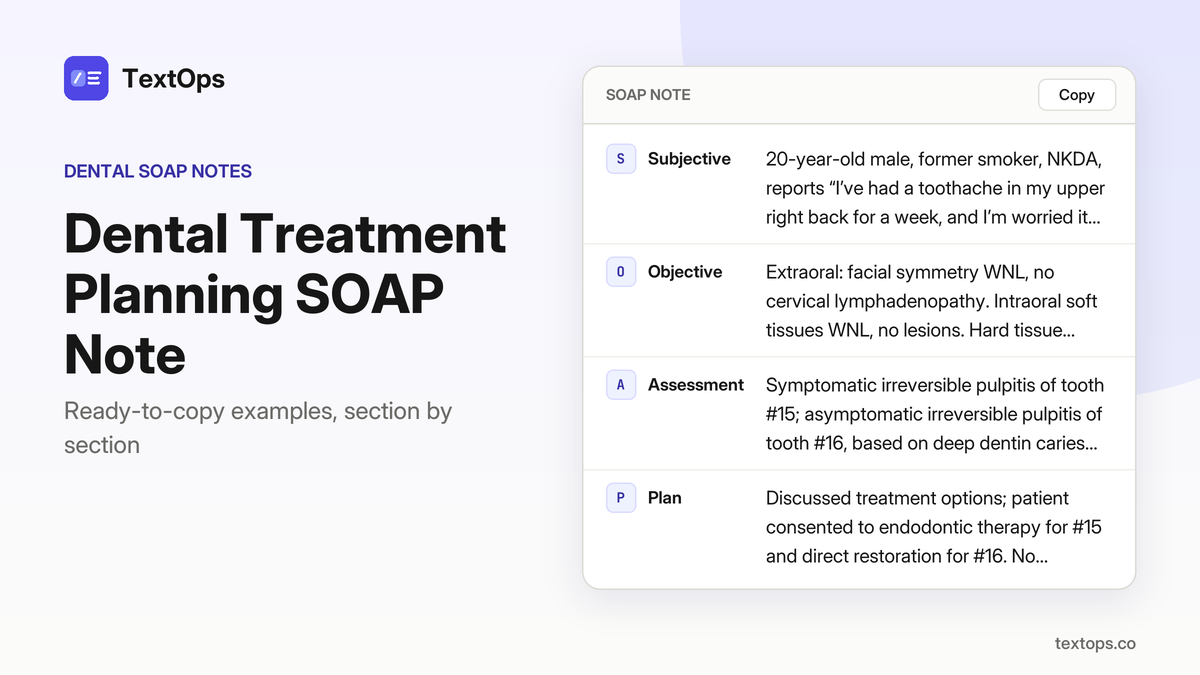
Fully formatted, copy-ready intraoral examination SOAP notes cover every common visit type. Notes are grouped by visit category and each template contains patient identification, objective exam findings, assessment, and plan. Quick-start tips highlight how to adapt a template for routine, emergency, or follow-up encounters, and mobile-friendly layout advice ensures seamless copy-paste on any device.
Routine & Initial Evaluations
Routine Check-Up, 45-Year-Old Male
SOAP note
SSubjective
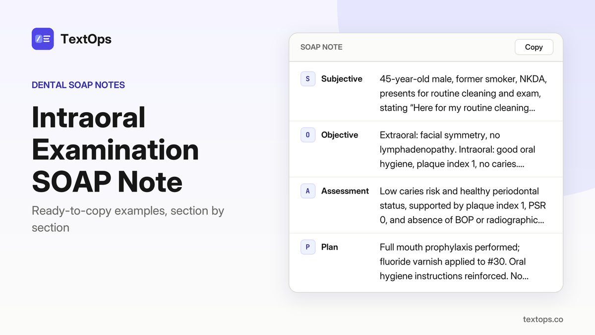
45-year-old male, former smoker, NKDA, presents for routine cleaning and exam, stating “Here for my routine cleaning and exam.”
OObjective
Extraoral: facial symmetry, no lymphadenopathy. Intraoral: good oral hygiene, plaque index 1, no caries. Periodontal: PSR 0 on all teeth, no BOP. Radiographs: bitewing series shows no new caries; periapical of #15 and #30 show no pathology.
AAssessment
Low caries risk and healthy periodontal status, supported by plaque index 1, PSR 0, and absence of BOP or radiographic pathology.
PPlan
Full mouth prophylaxis performed; fluoride varnish applied to #30. Oral hygiene instructions reinforced. No anesthesia or medications given. Next visit scheduled for routine check-up in 12 months.
First-Time Patient, 30-Year-Old Female
SOAP note
SSubjective
30-year-old female, type 2 diabetes on metformin, NKDA, first-time patient, “I have never been to a dentist before, I want a check-up.” HPI: no pain, routine check-up. PMH: HbA1c 7.8%.
OObjective
Extraoral: no swelling, TMJ normal. Intraoral: mild gingivitis on lower incisors, plaque index 2. Periodontal: PSR 1 on teeth #7-#10, no BOP. Radiographs: full-mouth series no caries; periapical of #16 shows slight radiolucency.
AAssessment
Gingivitis with moderate caries risk; periapical radiolucency on #16 likely early bone loss related to diabetes.
PPlan
Prophylaxis performed; scaling of lower incisors; fluoride varnish applied to #16; patient educated on glycemic control and oral hygiene; no anesthesia used; next visit: recall in 6 months for maintenance.
First-Time Patient with Burning Mouth Syndrome
SOAP note
SSubjective
28-year-old male, seasonal allergies, NKDA; CC: “My mouth feels like it’s on fire, especially when I eat spicy food”; HPI: burning sensation localized to buccal mucosa, worsened by spicy foods, no pain with cold or heat, no swelling, no systemic symptoms; PMH: seasonal allergies, no medications.
OObjective
Extraoral: no swelling, no lymphadenopathy; Intraoral: buccal mucosa erythematous, no discrete lesions; Periodontal: PSR 0, no BOP; Radiographs: bitewing series normal; Systemic signs: none.
AAssessment
Burning mouth syndrome, supported by erythematous buccal mucosa, absence of lesions or pathology on radiographs, and typical symptom pattern.
PPlan
Referred to oral medicine for further evaluation; advised to avoid irritants, follow a bland diet, maintain oral hygiene; no drugs prescribed; next visit: follow-up with oral medicine in 2 weeks.
Second Opinion for Complex Root Caries
SOAP note
SSubjective
67-year-old male, hypothyroidism on levothyroxine, penicillin allergy (rash); chief complaint: “I have a deep cavity on my lower right first molar and it hurts when I bite”; HPI: pain increases with biting, no fever or swelling; PMH: controlled hypothyroidism; no other drug allergies; no systemic signs.
OObjective
Extraoral: no swelling, no lymphadenopathy; Intraoral: extensive caries on tooth #30, deep into outer dentin, no pulpal exposure, no swelling; Periodontal: PSR 1 on #30, no BOP; Radiographs: periapical of #30 shows radiolucency extending to the root apex, no periapical abscess; Anesthesia: 2% lidocaine with 1:100,000 epinephrine, 1.8 mL (1 cartridge), IANB, aspiration negative.
AAssessment
Symptomatic irreversible pulpitis with symptomatic apical periodontitis, supported by deep caries, radiolucency to apex, and pain on biting.
PPlan
Endodontic therapy planned; informed consent obtained; antibiotic not indicated due to lack of systemic signs; no drugs prescribed; patient instructed on oral hygiene, pain control with NSAIDs if needed; next visit: endodontic treatment completion.
Emergency & Acute Pain Episodes
Emergency, 60-Year-Old Male with Toothache
SOAP note
SSubjective
60-year-old male with controlled hypertension on lisinopril, NKDA, reports “My lower right molar has been throbbing for the last 48 hours and it hurts when I bite down.” HPI: 48-hour throbbing pain, exacerbated by biting, no systemic symptoms. PMH: hypertension, no allergies.
OObjective
Tooth #30 shows deep enamel caries extending into outer dentin, no restoration; palpation of buccal cortical plate reveals localized swelling 4 mm in diameter; provisional probing depth 6 mm on distal surface with bleeding on probing; extraoral exam: no cervical lymphadenopathy, trismus absent; temperature 98.6 °F.
AAssessment
Symptomatic irreversible pulpitis with symptomatic apical periodontitis, supported by deep caries, 6 mm probing depth, bleeding on probing, and localized swelling.
PPlan
Administered 2% lidocaine with 1:100,000 epinephrine, 1.8 mL (1 cartridge), IANB; performed pulpotomy with calcium hydroxide dressing and placed a temporary stainless steel crown; prescribed ibuprofen 400 mg q6h PRN up to 3 days and acetaminophen 500 mg q6h PRN up to 3 days; instructed on pain control, oral hygiene, and to return in 7 days for definitive root canal.
Dental Trauma, 15-Year-Old Male
SOAP note
SSubjective
15-year-old male, allergic to penicillin (rash); chief complaint: “I fell in the playground and my front tooth is knocked out and I have a sharp pain when I bite.” History of present illness: trauma, no other symptoms.
OObjective
Extraoral: facial symmetry intact, no lymphadenopathy, temperature 98.4 °F. Intraoral: tooth #7 avulsed, socket shows mild bleeding, no fracture of adjacent teeth, no swelling on palpation.
AAssessment
Tooth avulsion with suspected pulpal necrosis; findings: avulsed tooth, mild bleeding, no swelling.
PPlan
Re-insertion of tooth #7 after 2% lidocaine infiltration, splinted with composite resin. Anesthesia used: 4% articaine with 1:100,000 epinephrine, 1.8 mL buccal infiltration. Prescribed chlorhexidine gluconate 0.12% 15 mL rinse for 30 seconds, twice daily for 7 days. Instructed to avoid hard foods, maintain oral hygiene, report bleeding or pain. Follow-up in 48 hours for vitality testing.
Dental Emergency, 30-Year-Old Female with Abscess
SOAP note
SSubjective
30-year-old female with asthma on albuterol PRN, NKDA, reports a swelling on the lower right side and a hot, throbbing pain that began this morning.
OObjective
Tooth #30 shows deep caries into outer dentin with pulpal exposure; buccal cortical plate swelling 6 mm, tender to palpation; probing depth 7 mm on distal surface with bleeding on probing; cervical lymphadenopathy present; temperature 99.2 °F; trismus mild, opening 35 mm.
AAssessment
Acute apical abscess with pulpal necrosis, evidenced by deep caries, pulpal exposure, 6 mm swelling, 7 mm probing depth, bleeding on probing, cervical lymphadenopathy, and low-grade fever.
PPlan
Administered 2% lidocaine with 1:100,000 epinephrine, 1.8 mL (1 cartridge) IANB, performed incision and drainage of the abscess, prescribed amoxicillin 500 mg TID for 7 days, ibuprofen 400 mg Q6H PRN up to 3 days, instructed on pain control, oral hygiene, and scheduled follow-up in 7 days.
Dental Emergency, 50-Year-Old Male with Severe Pain
SOAP note
SSubjective
50-year-old male, taking low-dose aspirin, NKDA; CC: “My upper left molar hurts so badly I can’t eat anything”; HPI: severe pain, no systemic signs; PMH: aspirin only.
OObjective
Tooth #16 shows extensive caries into outer dentin, no restoration; buccal cortical plate swelling 5 mm, tender; probing depth 5 mm mesial, BOP present; extraoral: no lymphadenopathy, temperature 98.7 °F, trismus absent.
AAssessment
Symptomatic irreversible pulpitis with symptomatic apical periodontitis, supported by deep caries, tender swelling, probing depth and bleeding.
PPlan
Administered 2% lidocaine with 1:100,000 epinephrine, 1.8 mL buccal infiltration; performed pulpectomy with calcium hydroxide dressing, placed temporary crown; prescribed ibuprofen 400 mg q6h PRN up to 3 days; instructed oral hygiene, avoid chewing on the side; scheduled definitive RCT in 7 days.
Emergency Visit for Acute Pericoronitis
SOAP note
SSubjective
44-year-old male, NKDA, presents with acute pericoronitis of tooth #32; says “My wisdom tooth is swollen and I can’t open my mouth fully.” HPI: swelling started 2 days ago, pain worsens with chewing, no fever. PMH: no significant medical history.
OObjective
Tooth #32 partially erupted, follicular space 6 mm; surrounding mucosa erythematous, edematous, 4 mm tender swelling; extraoral: no lymphadenopathy, T 98.5 °F; trismus 30 mm opening.
AAssessment
Acute pericoronitis of tooth #32, supported by partial eruption, follicular space >5 mm, erythematous mucosa, tender swelling, and trismus.
PPlan
Administered 2% lidocaine with 1:100,000 epinephrine, 1.8 mL IANB; performed incision and drainage of pericoronal flap; prescribed chlorhexidine gluconate 0.12% 15 mL rinse, 30 seconds, twice daily for 7 days; instructed on oral hygiene, scheduled follow-up in 7 days.
Root Surface Caries, 50-Year-Old Male
SOAP note
SSubjective
50-year-old male, smoker 5 cigarettes/day, NKDA, reports “My lower right molar feels gritty and I have a dull ache when I bite.” History: pain present for 2 days, no fever or swelling.
OObjective
tooth #30 shows root surface caries on distal surface extending into outer dentin; provisional probing depth 4 mm on distal surface, bleeding on probing; extraoral: no lymphadenopathy, temperature 98.6 °F, trismus absent.
AAssessment
root surface caries with symptomatic irreversible pulpitis, supported by caries depth, PD 4 mm, BOP, and pain on biting.
PPlan
administered 2% lidocaine with 1:100,000 epinephrine, 1.8 mL (1 cartridge), IANB; performed selective caries removal to dentin; placed glass ionomer restoration; prescribed ibuprofen 400 mg every 6 hours PRN, up to 3 days; instructed to avoid hard foods, maintain oral hygiene; next visit in 14 days for restoration check.
Dental Prosthesis, 70-Year-Old Female
SOAP note
SSubjective
70-year-old female, former smoker, NKDA, reports “My upper left partial denture keeps slipping and I feel pain when I bite.”
OObjective
Extraoral: no lymphadenopathy, temperature 98.4 °F, trismus absent. Intraoral: upper left partial denture shows poor fit with undercuts on mucosa, no tooth mobility; mucosal ulceration noted at denture contact sites.
AAssessment
Prosthetic irritation with mucosal ulceration, supported by poor denture fit and ulcerative mucosal changes.
PPlan
Removed partial denture, cleaned mucosa, fabricated new custom tray; scheduled impression taking in 3 days; instructed on denture hygiene; no drugs administered.
Post-Extraction & Trauma Follow-Ups
Post-Extraction Follow-Up, Day 3
SOAP note
SSubjective
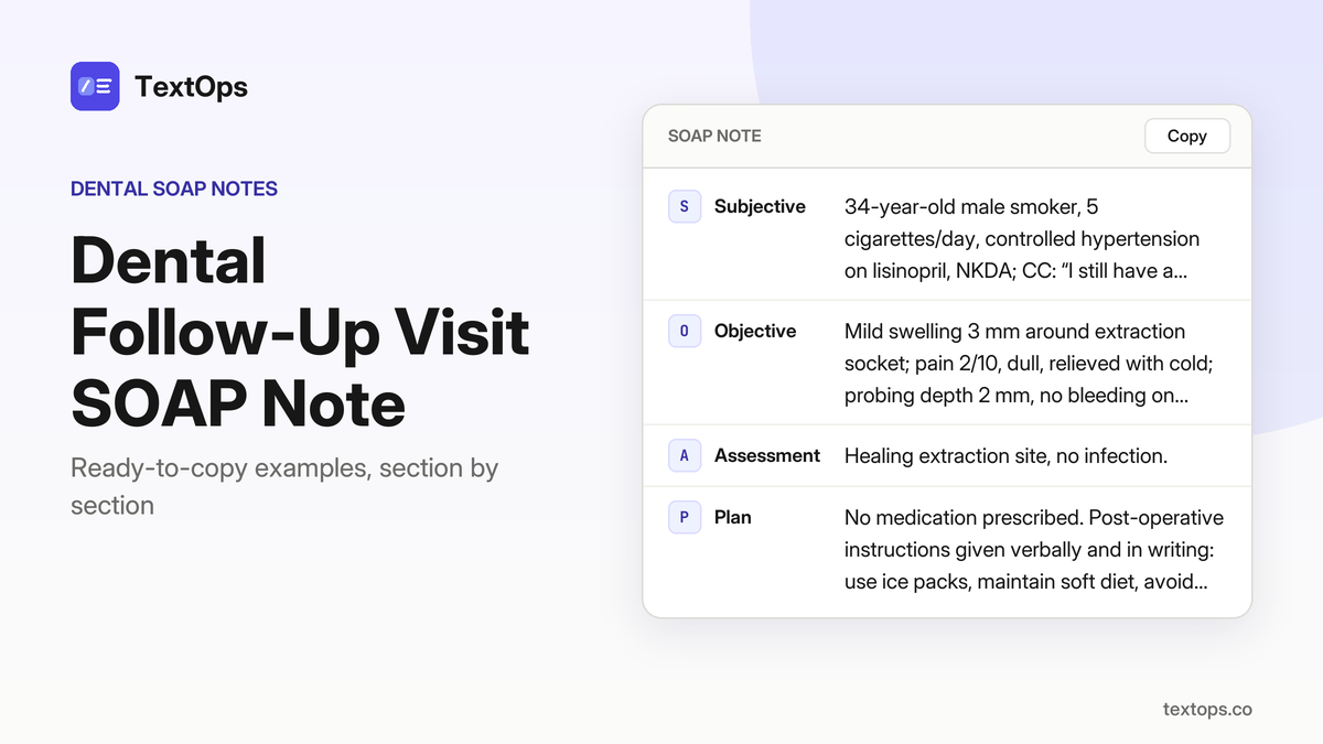
62-year-old male with type 2 diabetes on metformin, NKDA, reports “I’m fine, just a bit sore.”
OObjective
Extraction site on #30 shows mild erythema, soft tissue intact, probing depth 2 mm, no bleeding on probing, no drainage, no swelling or trismus; temperature 98.6 °F; patient reports mild discomfort 2/10; no lymphadenopathy.
AAssessment
Healing post-extraction, no infection, supported by erythema only, clear probing, absence of drainage or swelling.
PPlan
Reinforced oral hygiene instructions, prescribed chlorhexidine gluconate 0.12 % rinse 15 mL twice daily for 7 days, advised to monitor for increased pain or drainage, next visit in 7 days for final healing assessment.
Dental Trauma, 8-Year-Old Female
SOAP note
SSubjective
8-year-old female, seasonal allergies, NKDA; CC: “My front tooth hurts when I bite.” HPI: pain 4/10 on biting, no swelling or systemic symptoms.
OObjective
Extraoral: soft tissue intact, no swelling; Intraoral: #7 shows a shallow enamel fracture labial surface, mobility grade 0, percussion non-tender; Probing depth 1 mm, BOP negative; Periodontal: PSR 0; Radiograph: none taken; Tests: none.
AAssessment
Enamel fracture of #7, no pulpal involvement.
PPlan
Composite restoration of #7 labial surface performed under 4% articaine 1:100,000 epinephrine, 1.8 mL infiltration (1 cartridge). POI: avoid hard foods for 24 h, apply cold packs for swelling, maintain oral hygiene, return if pain worsens or swelling develops.
Implant & Surgical Follow-Ups
Dental Implant Surgery, 45-Year-Old Male
SOAP note
SSubjective
45-year-old male, hypothyroidism on levothyroxine, allergic to penicillin (rash); chief complaint: “I have a sore spot where the implant was placed”; no other systemic signs.
OObjective
Extraoral: no swelling, trismus, or lymphadenopathy; temperature 98.6 °F. Intraoral: implant site #20 shows healthy mucosa, 2 mm probing depth, no bleeding on probing. Radiograph: periapical shows good osseointegration, no radiolucency.
AAssessment
Implant site healing, no infection.
PPlan
Anesthesia: 2% lidocaine with 1:100,000 epinephrine, 1.8 mL (1 cartridge), IANB. Intra-operative irrigation with saline. Post-operative instructions given. Prescribed ibuprofen 400 mg PO q6h PRN for 3 days. Next visit: routine follow-up in 2 weeks.
Dental Implant Maintenance, 55-Year-Old Male
SOAP note
SSubjective
55-year-old male, controlled hypertension on lisinopril, NKDA, reports “Just a routine check-up for my implant.”
OObjective
Extraoral normal, intraoral mucosa pink around #15, no recession, probing depth 1 mm, no pain or swelling, temperature 98.7 °F, no lymphadenopathy; periapical radiograph shows stable implant, smooth surface, no bone loss.
AAssessment
Stable implant, no peri-implant disease, supported by 1 mm probing depth, healthy mucosa, and radiographic bone level.
PPlan
Cleaned implant surface with ultrasonic scaler and Polident brush, reinforced oral hygiene instructions, no drugs prescribed, next maintenance scheduled in 6 months.
Dental Implant Surgery, 65-Year-Old Female
SOAP note
SSubjective
65-year-old female, no significant medical history, states “I need a new implant in the back of my mouth”; temperature 98.4 °F, no lymphadenopathy.
OObjective
Extraoral normal; intraoral CBCT shows 10 mm bone height, 8 mm width at #30, no sinus involvement or infection; periapical radiograph shows healthy bone; no swelling; IANB anesthesia 2% lidocaine with 1:100,000 epinephrine, 1.8 mL.
AAssessment
Indication for implant placement in #30, supported by adequate bone dimensions and absence of infection.
PPlan
Placed 4.0 mm × 10 mm implant with primary stability 35 Ncm, sutured; prescribed ibuprofen 400 mg PO q6h PRN for 3 days; gave postoperative instructions and scheduled follow-up in 2 weeks for suture removal and healing assessment.
Dental Implant Failure, 65-Year-Old Female
SOAP note
SSubjective
65-year-old female with asthma, NKDA, reports “the implant in my back tooth keeps coming loose”; history of asthma managed with albuterol as needed.
OObjective
Extraoral normal; intraoral shows mucosal recession over #30, probing depth 6 mm, bleeding on probing; periapical radiograph reveals radiolucency around the implant apex; no swelling, temperature 98.9 °F, no lymphadenopathy.
AAssessment
Peri-implantitis with implant failure, evidenced by deep probing, BOP, and radiolucency around the implant apex.
PPlan
Removed implant, curetted bone, placed antibiotic-laden bone graft, sutured; prescribed amoxicillin 500 mg PO q8h for 7 days and chlorhexidine 0.12 % rinse 15 mL twice daily; instructed to maintain oral hygiene, avoid trauma, and return in 2 weeks for healing assessment.
Re-Evaluation of Implant Site After Bone Graft
SOAP note
SSubjective
64-year-old male, chief complaint: “Checking the graft site after my implant.” Medical history: daily low-dose aspirin, NKDA.
OObjective
Extraoral normal; intraoral mucosa healthy, no erythema, probing depth 2 mm, BOP negative. Periodontal: PD 2 mm, no bleeding. Radiographic: periapical view shows graft integration, no radiolucency. No swelling, temperature 98.5 °F, no lymphadenopathy.
AAssessment
Successful bone graft, implant site healing supported by healthy mucosa, shallow probing depth, and radiographic evidence of integration.
PPlan
No intervention performed; reinforced oral hygiene instructions given. Patient advised to maintain plaque control and avoid trauma to site. Next visit scheduled for implant placement in 4 weeks.
Post-Surgical Follow-Up for Maxillary Implant
SOAP note
SSubjective
24-year-old male, no significant medical history, NKDA, reports “Just checking on my new upper implant”; no pain or swelling, temperature 98.6°F, no lymphadenopathy.
OObjective
Extraoral normal; intraoral soft tissue pink, probing depth 1 mm, no bleeding on probing; periodontal screening shows healthy mucosa; periapical radiograph shows good osseointegration, no bone loss; no swelling, temperature normal.
AAssessment
Stable maxillary implant, no complications, based on pink mucosa, 1 mm probing depth, absence of bleeding, and radiographic osseointegration.
PPlan
Cleaned implant site, reinforced hygiene instructions, no drugs prescribed; next maintenance visit scheduled in 6 months.
Dental Implant Evaluation, 40-Year-Old Female
SOAP note
SSubjective
40-year-old female patient, smokes 5 cigarettes daily, NKDA, reports “I want to know if I can get an implant for my missing tooth.” HPI: no pain, no swelling, normal temperature 98.7 °F, no lymphadenopathy.
OObjective
CBCT shows 9 mm bone height, 7 mm width, no sinus involvement; periapical radiograph shows healthy bone, no pathology; extraoral and intraoral exam WNL; no swelling; temperature 98.7 °F; no lymphadenopathy.
AAssessment
Implant placement feasible in tooth #15 based on bone dimensions and absence of pathology.
PPlan
Discussed implant options, obtained informed consent, scheduled surgical placement in 2 weeks. No anesthesia or medications administered today. No postoperative instructions needed at this visit.
Sealant & Restoration Follow-Ups
Dental Sealant Removal, 9-Year-Old Male
SOAP note
SSubjective
9-year-old male, former smoker, NKDA, reports “I have a spot on my back tooth that looks like a black line”; history notes that the spot is on the buccal surface of tooth #30 and has been present for several weeks; no pain or sensitivity reported.
OObjective
extraoral normal; intraoral shows tooth #30 with intact enamel, no caries; buccal sealant partially intact as a thin film; no percussion or palpation sensitivity; oral hygiene good, plaque score 20 %; no lesions elsewhere.
AAssessment
Dental sealant retention on tooth #30, no caries; findings support that the thin film is a residual sealant without decay.
PPlan
Removed remaining sealant with hand scaler, cleaned surface, applied fluoride varnish; no anesthesia used; instructed patient and parent on maintaining plaque control and to return in 6 months for sealant integrity check.
Dental Crowning, 30-Year-Old Male
SOAP note
SSubjective
30-year-old male, type 2 diabetes on metformin, NKDA; chief complaint: “My front tooth is cracked and hurts when I bite”; pain localized to biting, no swelling or fever; past medical history: diabetes, medication: metformin; no known drug allergies.
OObjective
Tooth #7 has a large composite restoration on the buccal surface with a fracture line extending to the pulp chamber; percussion is tender and bite pain is present; periodontal probing shows PD 2 mm, BOP negative; periapical radiograph shows the restoration intact with no periapical radiolucency.
AAssessment
Fractured crown with pulpal involvement, symptomatic irreversible pulpitis—fracture line to pulp, tender percussion, bite pain, and absence of periapical pathology support the diagnosis.
PPlan
Local anesthesia: 2% lidocaine with 1:100,000 epinephrine, 1.8 mL buccal infiltration; pulpectomy performed, root canals obturated, post and core placed, full-coverage crown fabricated and cemented; postoperative instructions: maintain oral hygiene, avoid hard foods, use fluoride toothpaste; next visit: crown fit check in 2 weeks.
Re-Evaluation of Dental Sealants After 6 Months
SOAP note
SSubjective
51-year-old female, seasonal allergies, NKDA, reports “My sealants look a bit worn, but I don't have any pain.”
OObjective
Tooth #19 buccal sealant shows visible wear but remains intact; no caries detected clinically or on the PA radiograph. Plaque index is 15%, indicating moderate plaque accumulation; no sensitivity on biting or percussion; periodontal probing shows PD 1 mm and BOP negative, confirming healthy periodontal tissues around the sealant.
AAssessment
Well-maintained sealant, no caries; the wear is superficial and does not compromise the sealant’s protective function.
PPlan
Reinforced sealant with light-cure resin applied to the buccal surface of #19; patient advised to continue twice-daily brushing, use fluoride toothpaste, and maintain regular dental visits; no medication prescribed; next visit scheduled in 6 months for routine sealant evaluation.
Routine Maintenance & Re-Evaluation
Periodontal Maintenance, 55-Year-Old Male
SOAP note
SSubjective
55-year-old male, hypothyroidism on levothyroxine, CC: “Here for my regular cleaning and check-up.” HPI: routine maintenance visit.
OObjective
Extraoral: WNL. Intraoral: plaque score 35%; probing depths 2 mm bilaterally on tooth #30, BOP on buccal surface; radiograph: no caries, no bone loss. Periodontal: Stage I periodontitis, Grade A.
AAssessment
Stage I periodontitis, Grade A, supported by 2 mm probing depths, BOP, and normal radiographic bone levels.
PPlan
Professional cleaning performed; scaling and root planing on tooth #30 buccal surface; oral hygiene instruction reinforced; chlorhexidine 0.12% rinse 15 mL, 30 seconds, twice daily for 7 days prescribed. Next visit in 6 months for maintenance.
Dental Anxiety Management, 38-Year-Old Female
SOAP note
SSubjective
38-year-old female, controlled hypertension on lisinopril, NKDA, reports anxiety about cleaning, “I have a bit of anxiety about the cleaning, but I’m here for my routine visit.”
OObjective
Probing depths 1 mm on all surfaces of tooth #15, BOP none, plaque 20 %; periapical radiograph shows no caries or bone loss; extraoral and intraoral otherwise normal.
AAssessment
Healthy periodontium on tooth #15, supported by 1 mm probing depths, absence of BOP, and radiographic bone levels.
PPlan
Routine prophylaxis performed; no anesthesia used; patient instructed in deep-breathing and music relaxation; advised to return in 6 months for next cleaning; no medications prescribed.
Dental Implant Failure, 60-Year-Old Male
SOAP note
SSubjective
60-year-old male, no significant medical history, reports “I’m here for my regular check-up and to discuss the implant failure I had last year.”
OObjective
Extraoral normal. Intraoral: implant at #20 shows peri-implant mucositis, probing depth 4 mm, BOP present. Periodontal screening: Stage I periodontitis, Grade A. Radiograph: mild bone loss around implant, no fracture.
AAssessment
Peri-implant mucositis, Stage I periodontitis, Grade A, supported by 4 mm probing depth, BOP, and radiographic bone loss.
PPlan
Debridement of implant surface performed. Chlorhexidine gluconate 0.12% 15 mL rinse, 30 seconds, twice daily for 14 days prescribed. Advised oral hygiene reinforcement and to return in 4 weeks for reassessment.
Dental Pain Management, 55-Year-Old Male
SOAP note
SSubjective
55-year-old male, asthma on albuterol PRN, NKDA, reports “I have a dull ache in my upper left canine that’s been there for a week.” He describes the pain as dull, 3/10, with no triggers and no systemic symptoms.
OObjective
Probing depth 3 mm on the buccal surface of tooth #11; bleeding on probing present; pain rating 3/10; periapical radiograph shows no caries and no periapical radiolucency.
AAssessment
Stage I periodontitis, Grade A, based on a 3 mm probing depth, BOP, and absence of radiographic pathology.
PPlan
Scaling and root planing performed on the buccal surface of tooth #11; oral hygiene instruction reinforced; no medications prescribed; patient advised to return in 6 months.
Dental Hygiene, 25-Year-Old Female
SOAP note
SSubjective
25-year-old female, NKDA, daily low-dose aspirin, chief complaint: “I’d like a routine cleaning and to check my teeth for any new decay.”
OObjective
Extraoral normal. Intraoral: tooth #22 shows probing depths 1 mm on all surfaces; no BOP; plaque index 15 %; radiograph shows no caries or bone loss. Periodontal screening: healthy periodontium.
AAssessment
Healthy periodontium, supported by probing depths ≤1 mm, absent BOP, and radiographic bone levels within normal limits.
PPlan
Prophylaxis performed with scaling and polishing; patient advised to brush twice daily with fluoride toothpaste, floss nightly, and use interdental brushes as needed. No medication prescribed. Next routine cleaning scheduled in 6 months.
Dental Pain Management, 60-Year-Old Female
SOAP note
SSubjective
60-year-old female, NKDA, reports “I have a sharp pain when I bite down on the upper right canine.” She denies other symptoms; medical history unremarkable.
OObjective
Extraoral: facial symmetry, no lymphadenopathy. Intraoral: tooth #6 shows a 2 mm probing depth on buccal surface, no BOP, no caries, radiograph reveals no periapical radiolucency. Periodontal screening: Stage I periodontitis, Grade A.
AAssessment
Stage I periodontitis, Grade A of tooth #6, supported by 2 mm probing depth, absence of BOP, and radiographic normalcy.
PPlan
Scaling and root planing performed on buccal surface of #6; patient advised to avoid hard foods for 48 hours. No drugs prescribed. Follow-up scheduled in 4 weeks for reassessment.
Recurrent Gingival Bleeding in a Smokeless Patient
SOAP note
SSubjective
71-year-old female, smokes 5 cigarettes daily, NKDA; chief complaint: “I keep bleeding when I brush my lower left premolars”; history: recurrent gingival bleeding during brushing, no pain, no swelling; medical history: none; medication: none; allergies: none.
OObjective
Extraoral: no facial asymmetry, lymph nodes non-tender; intraoral: plaque index 40%, BOP present on all surfaces of tooth #20; periodontal: probing depth 3 mm buccally on #20, BOP on all surfaces; radiograph: mild bone loss around #20, no caries; tests: none.
AAssessment
Stage II periodontitis, Grade B, supported by 3 mm probing depth, BOP on all surfaces, mild bone loss, and 40% plaque.
PPlan
Professional cleaning performed; scaling and root planing on tooth #20; patient advised to quit smoking, instructed to use chlorhexidine gluconate 0.12% rinse 15 mL, 30 seconds, twice daily for 14 days; next visit scheduled in 4 weeks.
Post-Orthodontic Retainer Check-Up
SOAP note
SSubjective
34-year-old male, former smoker, NKDA, presents with chief complaint: “I’d like to check my retainer and see if it’s still fitting properly.” History: routine maintenance visit, no pain, no swelling, no systemic signs.
OObjective
Retainer: clear acrylic, well-fitted, no gaps or fractures; Oral hygiene: adequate, no plaque accumulation; Radiograph: no caries, no periodontal changes; Extraoral: facial symmetry, no lymphadenopathy, TMJ normal.
AAssessment
No active pathology; retainer fitting acceptable.
PPlan
Retainer inspection and minor adjustment performed; patient instructed to clean retainer daily with mild soap; no drugs prescribed; scheduled next check-up in 6 months.
Follow-Up After Endodontic Retreatment
SOAP note
SSubjective
50-year-old male, type 2 diabetes on metformin, NKDA, reports “I’m here for my routine cleaning and to discuss my diabetes management with oral health.”
OObjective
Extraoral examination shows no facial asymmetry, no lymphadenopathy, and normal TMJ function. Intraoral inspection of tooth #16 reveals probing depths of 2 mm on all surfaces, no bleeding on probing, and a plaque index of 20 %. No carious lesions or restorations are present, and the periapical radiograph demonstrates intact bone levels with no radiolucencies or root resorption. Periodontal charting confirms a healthy periodontium with no pockets deeper than 3 mm.
AAssessment
Healthy periodontium; probing depths ≤3 mm, no BOP, radiographs normal, consistent with optimal periodontal health in the context of well-controlled type 2 diabetes.
PPlan
Prophylaxis performed with ultrasonic scaling and polishing; patient received counseling on the importance of glycemic control for periodontal maintenance; no medications prescribed; next cleaning scheduled in 6 months.
Restorative Procedures
Root Canal Therapy, 35-Year-Old Male
SOAP note
SSubjective
35-year-old male, seasonal allergies, NKDA, chief complaint: “I’ve had a sharp, throbbing pain in my lower right back tooth for the last week”; history: pain started a week ago, no swelling or trismus; medical history: seasonal allergies, no other conditions; medications: none; allergies: NKDA.
OObjective
Tooth #30 shows deep caries extending to outer dentin with pulpal exposure; periapical radiograph reveals a 3 mm radiolucency; probing depth 6 mm with BOP; extraoral exam normal; temperature 98.6 °F; anesthesia: 2% lidocaine with 1:100,000 epinephrine, 1.8 mL, IANB, aspiration negative.
AAssessment
Symptomatic irreversible pulpitis with acute apical periodontitis, supported by pulpal exposure, deep caries, radiolucency, deep probing depth, and BOP.
PPlan
Root canal therapy performed on #30: canal cleaned, shaped, irrigated with 2.5% NaOCl, obturated with gutta-percha and AH-Plus sealer; temporary restoration placed; no medications prescribed; instructed to avoid hard foods, maintain oral hygiene, return in 2 weeks for permanent restoration.
Orthodontic Referral, 12-Year-Old Female
SOAP note
SSubjective
12-year-old female, hypothyroidism on levothyroxine, allergic to penicillin (rash). Chief complaint: “My front teeth look crooked and I want them straight.” History: mild crowding of upper centrals, 4 mm overjet, no pain, no caries, no periodontal disease. PMH: levothyroxine, penicillin allergy.
OObjective
Extraoral: facial symmetry WNL, no swelling. Intraoral: upper central incisors mild crowding, overjet 4 mm, no caries, no periodontal pockets, BOP absent. Periodontal: PSR 0. Radiograph: panoramic shows all permanent teeth, no impactions, no root resorption. Tests: none.
AAssessment
Dental malocclusion – mild crowding, Class I malocclusion, requiring palatal expansion.
PPlan
Referred to orthodontist for comprehensive evaluation and treatment planning. Informed patient and parents of referral process, consent obtained verbally. No drugs administered. Next step: await orthodontic referral appointment.
Dental Sealant Placement, 7-Year-Old Male
SOAP note
SSubjective
7-year-old male, NKDA, controlled hypertension on lisinopril, chief complaint: “My dad says my molars look a bit rough.” HPI: no pain, routine check-up; PMH: hypertension, no allergies.
OObjective
Extraoral: normal. Intraoral: tooth #12 and #13 with smooth occlusal surfaces, no caries; probing depth 1 mm, no BOP; caries risk high due to early childhood caries history; radiograph shows no decay.
AAssessment
High caries risk, no active decay; supports sealant placement to prevent future caries.
PPlan
Applied 5 % fluoride sealant to occlusal surfaces of #12 and #13; no anesthesia used. Instructed parents on brushing technique and use of fluoride toothpaste. No drugs prescribed. Next visit: routine recall in 6 months.
Cavity Restoration, 28-Year-Old Male
SOAP note
SSubjective
28-year-old male, “There’s a deep cavity on my front tooth that hurts when I bite,” no significant medical history, allergic to penicillin (rash), temperature 98.7 °F.
OObjective
Tooth #7 shows class II caries on the mesiolingual surface extending to outer dentin, no pulpal exposure; probing depth 2 mm, no BOP; PA radiograph shows no periapical pathology; infiltration anesthesia 2% lidocaine with 1:100,000 epinephrine, 1.8 mL (1 cartridge), aspiration negative.
AAssessment
Reversible pulpitis, evidenced by deep caries to outer dentin, pain on biting, normal probing depth, no radiographic pathology, and absence of pulpal exposure.
PPlan
Prepared cavity, placed a 3 mm composite restoration using an incremental technique, finished and polished; no medications prescribed; instructed to maintain oral hygiene, avoid hard foods for 24 h, and return if pain persists.
Dental Crowning, 42-Year-Old Female
SOAP note
SSubjective
42-year-old female, asthma on albuterol PRN, NKDA, reports “My front tooth is cracked and I want it fixed”; tooth #7 shows a large fracture from incisal to cervical third with pulpal exposure, 3 mm probing depth, BOP present, no periapical pathology on PA, temperature 98.5 °F.
OObjective
Extraoral normal; intraoral tooth #7 fractured with exposed pulp, 3 mm probing depth, BOP; periodontal screening PSR 2; radiograph shows no periapical lesion; anesthesia 2% lidocaine with 1:100,000 epinephrine, 1.8 mL infiltration, aspiration negative.
AAssessment
Pulpal necrosis with symptomatic apical periodontitis, supported by pulpal exposure, BOP, and 3 mm probing depth.
PPlan
Root canal therapy completed on #7; composite core placed, followed by full-coverage porcelain-veneered crown; patient instructed to avoid hard foods for 24 h, use soft diet, maintain oral hygiene, and return in 2 weeks for follow-up to assess crown fit and healing.
Dental Sealant Removal, 7-Year-Old Female
SOAP note
SSubjective
7-year-old female patient, NKDA, takes low-dose aspirin daily, chief complaint: “My dad says the sealant on my molar is loose,” patient reports no pain or sensitivity, no history of decay in that tooth.
OObjective
tooth #3, occlusal surface partially detached sealant, no caries detected, probing depth 1 mm, no bleeding on probing, radiograph shows intact enamel and no periapical pathology, oral temperature 98.4 °F, patient appears comfortable.
AAssessment
sealant retention failure, no active decay, diagnosis confirmed by clinical and radiographic findings.
PPlan
removed old sealant with hand instruments, cleaned the occlusal surface with a rubber cup and pumice, applied a new 5 % fluoride sealant using a hand-held brush, no anesthesia required, instructed parent to avoid hard foods for 24 h, advised to brush twice daily with fluoride toothpaste, schedule a follow-up in 6 months to assess sealant integrity.
Dental Anxiety Assessment Before Simple Extraction
SOAP note
SSubjective
55-year-old female, no significant medical history, NKDA; chief complaint: “I’m nervous about having my tooth pulled”; history of present illness: anxiety level 8/10 on VAS, no swelling, trismus, or fever, temperature 98.6 °F.
OObjective
Tooth #30 shows large caries with pulpal exposure and a 4 mm periapical radiolucency; probing depth 6 mm, bleeding on probing present; patient reports high anxiety, no systemic signs.
AAssessment
Symptomatic irreversible pulpitis with acute apical periodontitis; patient anxiety.
PPlan
Conducted Modified Dental Anxiety Scale assessment; discussed extraction versus root canal; patient elected extraction with local anesthesia; scheduled extraction for next visit; no drugs prescribed today.
Adapting the SOAP Note for Different Visit Types
Begin each SOAP note with a concise, visit-specific objective focus: routine visits demand a full chart; emergencies require a problem-focused exam; post-extraction visits center on socket healing; maintenance visits emphasize periodontal status.
Routine & Initial Evaluations
Extraoral: facial symmetry, TMJ function, lymph nodes.
Intraoral: full charting of restorations, caries, periodontal probing, occlusion.
Radiographs: PA or BWX per risk assessment.
Assessment: baseline oral health, caries risk, periodontal status.
Plan: preventive measures, recall interval, patient education.
Emergency & Acute Pain Episodes
Extraoral: limited to trauma or swelling assessment.
Intraoral: focused exam on affected area, palpation, percussion, mobility.
Radiographs: targeted PA or CBCT if indicated.
Assessment: acute pathology (abscess, fracture, trauma).
Plan: immediate treatment (drainage, extraction, stabilization), pain control, antibiotic if systemic signs.
Maintenance & Re-Evaluation Visits
Extraoral: routine check of TMJ, facial symmetry.
Intraoral: periodontal charting, caries surveillance, restoration integrity.
Radiographs: periodic BWX or PAN as per protocol.
Assessment: disease control, progression.
Plan: scaling, polishing, sealant placement, recall interval.
Checklist for quick adaptation
Add extraoral findings only for trauma or swelling.
Limit intraoral exam to affected quadrant in acute pain.
Include socket assessment and POI for post-extraction.
Expand periodontal charting for maintenance visits.
Adjust radiograph type to visit purpose (targeted PA vs full-mouth).
Tailor assessment language to problem focus versus baseline status.
Keep subjective CC and HPI consistent across all visits.
Understanding Exam Scope: Limited vs Comprehensive
Limited intraoral exams focus on a single chief complaint. History is brief: the patient’s pain, onset, character, and any relevant medical conditions. The exam is confined to the affected quadrant or tooth, with targeted palpation, percussion, and mobility checks. Documentation records only findings that inform the immediate problem and a plan that addresses that issue alone. For example, a 60-year-old male with a pulpal abscess in tooth #36: record HPI, limited exam of #36, PA radiograph, diagnosis of acute apical abscess, and plan for extraction or root canal.
Comprehensive exams cover the entire mouth and are performed for new patients, after a long recall interval, or when systemic changes occur. History is full: PMH, medications, allergies, risk factors, and a detailed dental history. The intraoral exam includes full charting of restorations, caries, periodontal probing, occlusion, and oral cancer screening. Documentation lists all findings, radiographs (PA, BWX, FMX or PAN as indicated), and a treatment plan that addresses all oral health issues. For example, a 45-year-old male at a first-time check-up: record full history, complete charting, PA and BWX, diagnosis of mild periodontitis and multiple carious lesions, and a plan for restorations, scaling, and recall in 6 months.
Scope
History Depth
Exam Breadth
Documentation
Limited
Focused on complaint
Targeted to affected area
Problem-specific findings, single radiograph, immediate plan
Comprehensive
Full medical and dental
Full charting, periodontal, occlusion, cancer screen
Complete findings, multiple radiographs, long-term plan
Examples
Limited: 30-year-old female with tooth #12 pain → limited exam, PA, plan for RCT.
Comprehensive: 55-year-old male returning after 3 years → full exam, FMX, periodontal chart, treatment plan for crowns, scaling, and recall.
Mobile-Friendly Navigation Tips
Keep the page header sticky so the “Intraoral Examination SOAP Note” title stays visible while scrolling.
Collapse each note category (Routine, Emergency, Follow-up, etc.) into a tap-to-expand panel; this reduces on-screen clutter and lets you focus on one visit type at a time.
Inside each expanded panel, place the template in a single-line code block; the block should be the entire note so it can be selected with one tap.
Enable a “Copy all” button directly beneath the code block; the button should trigger a single-tap copy to clipboard, bypassing the need to drag text.
Add a small “Copied!” toast that disappears after a second, confirming the action without interrupting workflow.
Ensure the layout uses responsive font sizes (≥14 px) and wide touch targets (≥44 px) so tapping is accurate on small screens.
Quick-Start Copy-Paste Workflow
Open the charting screen, place cursor in the SOAP note field, and tap “Paste.”
Locate the desired template in the “Routine & Initial Evaluations” panel.
Tap the copy icon beside the code block, then confirm the clipboard copy.
Return to the chart, paste the text; the note will appear with placeholder tags.
Replace each placeholder—{patient name}, {age}, {CC}, {HPI}, {PMH}, {Allergies}—with the current patient’s data.
Verify the objective section for correct tooth numbers and findings; adjust if the exam differs.
Save the entry, sign, and schedule the next visit per the plan line.
These examples show structure and wording only. Record what you actually found and did, and follow your practice’s documentation requirements.
Dr. Rahul Joshi is a Specialist Orthodontist with extensive clinical experience in orthodontics, dentofacial orthopaedics, and dental aesthetics. Holding a Master of Dental Surgery (MDS) in Orthodontics and Dentofacial Orthopaedics and a Bachelor of Dental Surgery (BDS) from Manipal Academy of Higher Education, he brings expertise in orthodontic treatment planning, teeth alignment, and Invisalign treatment. With professional experience across India and the Netherlands, Dr. Joshi combines clinical knowledge with practical experience in orthodontic care, dental aesthetics, and improving patients' oral health.
Master of Dental Surgery (MDS) in Orthodontics and Dentofacial Orthopaedics — Manipal Academy of Higher Education
Bachelor of Dental Surgery (BDS) — Manipal Academy of Higher Education
Stop retyping your templates
Save any template to TextOps once, then insert it anywhere on the web without retyping. Share it with your team so everyone uses the same wording.