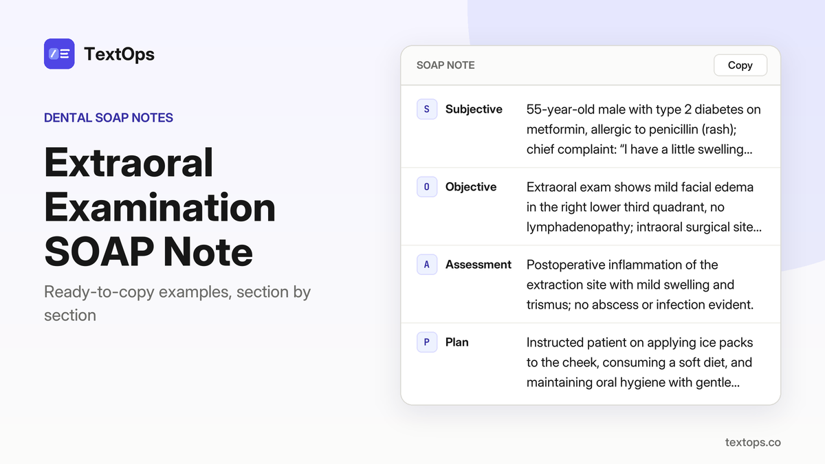
A ready-made, copy-ready SOAP note captures every element of a treatment-planning visit in a single, mobile-friendly layout. The note includes concise guidance on documenting informed consent, distinguishing a treatment-planning SOAP from a routine exam, and adapting the content to paper, EHR, or mobile charting systems. Each section is written for immediate use, with no extra narrative, ensuring accurate, contemporaneous documentation.
Initial Comprehensive Treatment Planning
First-Visit Adult with Moderate Caries
SOAP note
SSubjective
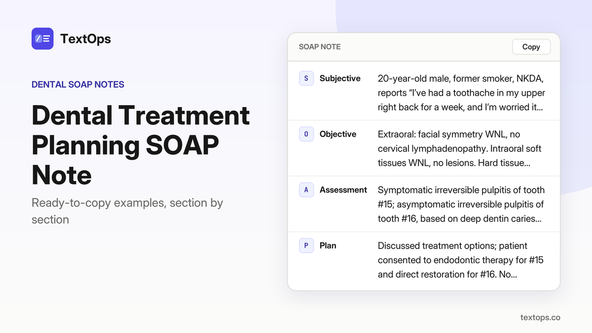
20-year-old male, former smoker, NKDA, reports “I’ve had a toothache in my upper right back for a week, and I’m worried it might be a cavity.” HPI: pain localized to #15, throbbing, worsens with chewing, no night pain. PMH: none.
OObjective
Extraoral: facial symmetry WNL, no cervical lymphadenopathy. Intraoral soft tissues WNL, no lesions. Hard tissue: caries on #15 mesial surface, deep dentin; caries on #16 occlusal surface, outer dentin; restoration on #14 Class II composite intact. Periodontal: PSR 1 on all teeth, no BOP. Radiographs: PA #15 shows proximal caries extending to outer dentin; PA #16 shows occlusal caries to outer dentin; no periapical radiolucency.
AAssessment
Symptomatic irreversible pulpitis of tooth #15; asymptomatic irreversible pulpitis of tooth #16, based on deep dentin caries, pain characteristics, and radiographic proximity to pulp.
PPlan
Discussed treatment options; patient consented to endodontic therapy for #15 and direct restoration for #16. No anesthesia administered today. Instructed on oral hygiene, diet, and follow-up in 1 week for endodontic appointment.
Follow-up and Recall Visits
Re-Evaluation of Root-Canal Therapy
SOAP note
SSubjective
74-year-old male, NKDA, reports “I still feel a little ache when I bite down”; no other symptoms.
OObjective
Intraoral exam shows tooth #15 restored with post and core, no caries, no mobility, mild BOP; probing PD 3 mm, CAL 0 mm; periapical radiograph shows resolved radiolucency, slight root resorption; temperature 98.6 °F, no swelling or lymphadenopathy.
AAssessment
Previously treated tooth #15, asymptomatic apical periodontitis; findings support resolved pathology with minimal residual inflammation.
PPlan
Reassured patient; no additional treatment indicated; instructed to maintain oral hygiene and report any new pain; scheduled for 12-month recall to monitor healing.
Periodontal Maintenance for Stage III Periodontitis
SOAP note
SSubjective
57-year-old male, controlled hypertension on lisinopril, NKDA, reports “My gums still bleed a little when I brush.”
OObjective
Extraoral: no facial swelling, lymph nodes not enlarged. Intraoral: generalized BOP, probing depths 5–6 mm in posterior sextants, CAL 4–5 mm; radiographs: bone loss up to 3/4 root in posterior teeth; periodontal charting: PSR 3 with furcation involvement grade I in tooth #18; BP 138/82 mmHg, HR 78 bpm.
AAssessment
Stage III periodontitis, Grade B, supported by generalized BOP, probing depths ≥5 mm, CAL 4–5 mm, radiographic bone loss 3/4 root, and PSR 3 with furcation grade I.
PPlan
Deep scaling and root planing of posterior sextants performed; oral hygiene instruction reinforced; chlorhexidine gluconate 0.12 % rinse 15 mL for 30 sec, twice daily prescribed; next visit in 3 months for maintenance.
Post-Extraction Healing Check – Day 3
SOAP note
SSubjective
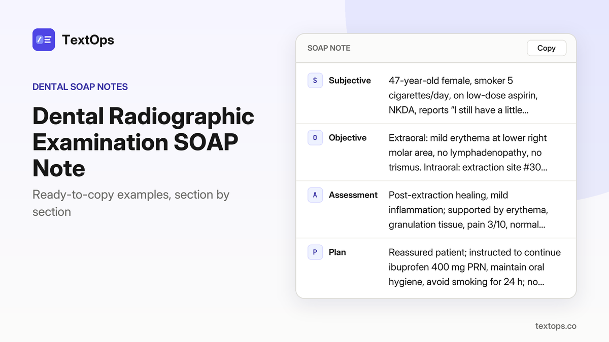
63-year-old male with type 2 diabetes on metformin, NKDA, reports “the socket feels a bit sore.” HPI: three days post-extraction of tooth #30, pain mild, no swelling. Medical history: diabetes, no other conditions.
OObjective
Extraoral: no facial swelling, no lymphadenopathy. Intraoral: extraction socket of #30 shows healthy granulation tissue, mild erythema, no suppuration. Probing: socket depth 3 mm, no bleeding on probing. Temperature 99.1 °F; blood glucose 145 mg/dL.
AAssessment
Healing extraction site #30, no infection, consistent with postoperative recovery.
PPlan
Advised to maintain oral hygiene, avoid vigorous rinsing, continue metformin. Prescribed ibuprofen 400 mg PO every 6 h PRN for pain. No anesthesia administered. Next visit: routine follow-up in 7 days to assess healing.
Periodontal Re-evaluation – 6-Month Recall
SOAP note
SSubjective
67-year-old male, chief complaint “My gums still bleed a little when I floss,” history of low-dose aspirin use, NKDA, no systemic symptoms.
OObjective
Extraoral exam shows no swelling or lymphadenopathy. Intraoral BOP present in 30 % of sites; probing depths 4–5 mm in molars, CAL 3–4 mm; PSR 2, no furcation involvement. Radiographs demonstrate bone loss stable compared to 6-month prior. Vital signs: BP 132/78 mmHg, HR 76 bpm.
AAssessment
Stage II Periodontitis, Grade A, supported by BOP, probing depths, CAL, and stable radiographic bone loss.
PPlan
Full-mouth scaling and root planing performed on molar sextants; oral hygiene reinforcement given; chlorhexidine gluconate 0.12 % rinse 15 mL for 30 s, twice daily prescribed; next visit scheduled in 3 months.
Periodontal Maintenance – 3-Month Recall
SOAP note
SSubjective
69-year-old female, chief complaint “My gums feel a bit swollen,” history of hypothyroidism on levothyroxine, allergic to penicillin (rash).
OObjective
Extraoral shows no facial swelling or lymphadenopathy; intraoral BOP in 25 % of sites, probing depths 3–4 mm in posterior sextants, CAL 2–3 mm; radiographs reveal unchanged bone levels; PSR 1, no furcation involvement; vital signs T 98.4 °F, BP 125/80 mmHg.
AAssessment
Stage I Periodontitis, Grade A, supported by BOP, shallow probing depths, minimal CAL, and stable radiographic bone levels.
PPlan
Performed supragingival scaling of all sextants; instructed on oral hygiene; prescribed chlorhexidine gluconate 0.12 % rinse 15 mL for 30 seconds, twice daily; next visit scheduled in 3 months.
Emergency and Acute Care
Emergency Extraction of Impacted Third Molar
SOAP note
SSubjective
58-year-old male, NKDA except penicillin rash, reports “My wisdom tooth is hurting and I can’t eat.” History: pain began 2 days ago, no fever or chills. O: impacted #32 with pericoronal radiolucency on PA, mild buccal vestibular swelling, no drainage, no systemic signs. A: Impacted third molar with pericoronitis, evidenced by radiolucency and clinical swelling. P: Administered 4% articaine 1:100,000, 1.8 mL IANB, aspiration negative; surgically extracted #32, sutured socket, prescribed ibuprofen 400 mg PO q6h PRN for 5 days, instructed on suture care and pain management, scheduled follow-up in 7 days.
Treatment of Acute Periapical Abscess
SOAP note
SSubjective
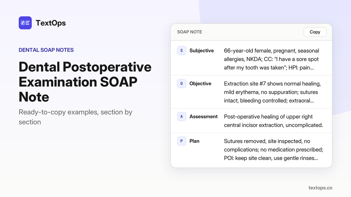
66-year-old male, seasonal allergies, NKDA, reports “sharp pain in my upper right first molar, especially when I bite.” He notes swelling in the buccal vestibule and a foul-smelling purulent discharge from tooth #3.
OObjective
Extraoral: mild facial swelling, palpable cervical lymphadenopathy; temperature 99.5 °F. Intraoral: buccal vestibular swelling, purulent drainage from #3. Periodontal: normal probing, no periodontal pockets. Radiograph: PA of #3 shows periapical radiolucency. Tests: none performed beyond visual inspection.
AAssessment
Acute apical abscess of tooth #3, supported by purulent drainage, periapical radiolucency, swelling, mild fever, and cervical lymphadenopathy.
PPlan
2% lidocaine with 1:100,000 epinephrine, 1.8 mL infiltration, aspiration negative; incision and drainage of the abscess performed. Prescribed amoxicillin 500 mg PO TID for 7 days. Instructed patient to avoid hard foods and report worsening pain or fever. Next visit: follow-up in 48 hours to assess drainage and pain control.
Emergency Pain Management – Toothache
SOAP note
SSubjective
29-year-old female patient, asthma on albuterol PRN, NKDA, reports “My front tooth hurts all the time, even when I drink cold.” HPI: pain constant, sharp, triggered by cold liquids, no swelling or drainage, no systemic symptoms.
OObjective
Extraoral: no facial swelling, lymphadenopathy, or trismus. Intraoral: deep enamel-dentin caries on facial surface of tooth #7, no abscess or drainage, no mobility, gingiva normal. Periodontal: probing depths within normal limits. Radiograph: periapical radiograph shows no periapical radiolucency.
AAssessment
Symptomatic irreversible pulpitis of tooth #7, supported by deep caries, pain with cold, and lack of periapical pathology.
PPlan
No local anesthesia administered. Prescribed ibuprofen 400 mg PO q6h PRN for 3 days for pain control. Advised oral hygiene, avoid extreme temperatures, and return for definitive endodontic treatment within 1–2 weeks.
Dental Emergency – Fracture of Incisor
SOAP note
SSubjective
51-year-old male, smoker 5 cigarettes/day, NKDA, reports “I broke my front tooth when I fell.” HPI: acute fracture of tooth #9, pain localized, no swelling, no drainage, no systemic symptoms.
OObjective
Extraoral: facial symmetry, no lymphadenopathy, no facial swelling. Intraoral: oblique fracture of #9 extending to pulp, no discoloration of surrounding teeth, no mobility. Periodontal: probing depths 2 mm, no BOP. Radiograph: PA of #9 shows fracture line to pulp, no periapical radiolucency.
AAssessment
Fractured tooth #9 with pulp exposure, consistent with oblique pulpal fracture on clinical and radiographic findings.
PPlan
Administered 2% lidocaine with 1:100,000 epinephrine, 1.8 mL infiltration, aspiration negative. Performed root canal therapy under local anesthesia. POI: maintain oral hygiene, avoid chewing on fractured side, use soft diet, return in 1 week for post-treatment evaluation.
Dental Emergency – Abscess Drainage
SOAP note
SSubjective
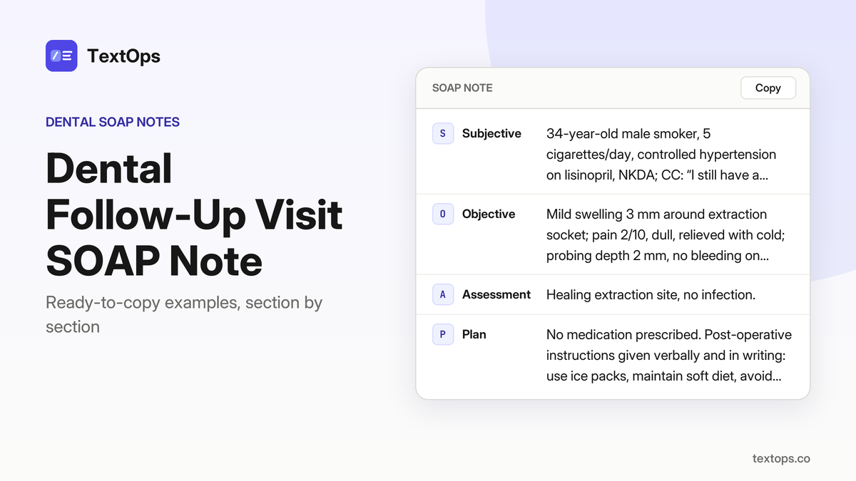
44-year-old male, former smoker, NKDA, reports “swelling on my lower right side, it’s painful.” He notes the swelling has been present for 2 days, worsens with chewing, and is accompanied by a throbbing ache; temperature 100.2 °F, trismus noted.
OObjective
Extraoral: palpable swelling in lower right vestibule; intraoral: purulent drainage from tooth #30; radiograph: PA shows periapical radiolucency; vital signs: T 100.2 °F, trismus present; anesthesia: 2% lidocaine with 1:100,000 epinephrine, 1.8 mL IANB, aspiration negative.
AAssessment
Acute apical abscess of tooth #30, supported by purulent drainage, periapical radiolucency, fever, and trismus.
PPlan
Incision and drainage performed under IANB; amoxicillin 500 mg PO TID for 7 days prescribed; patient instructed to take full course, maintain oral hygiene, avoid hard foods, and return if pain worsens or swelling recurs. Next visit scheduled in 7 days for reassessment.
Surgical and Implant Procedures
Crown Placement for Endodontically Treated Tooth
SOAP note
SSubjective
28-year-old male, NKDA, reports “my tooth has been hurting for a week and I need a crown.” He has no systemic signs.
OObjective
Root canal treatment completed on #15; 3 mm of remaining tooth structure; missing full-coverage restoration; PA shows no periapical radiolucency; PD 2 mm; BOP negative; infiltration anesthesia 2% lidocaine with 1:100,000 epinephrine, 1.8 mL.
AAssessment
Previously treated tooth #15, asymptomatic apical periodontitis, supported by intact periapical tissues and probing depth within normal limits.
PPlan
Crown preparation on #15, porcelain-veneered crown placed and cemented with resin cement; post-op instructions given; ibuprofen 400 mg q6h PRN up to 3 days, acetaminophen 500 mg q6h PRN up to 3 days; next visit in 1 week for restoration check.
Full-Mouth Periodontal Surgery
SOAP note
SSubjective
47-year-old female, controlled hypertension on lisinopril, NKDA, reports “My gums bleed a lot and I have bad breath.” HPI: bleeding on probing, halitosis, no pain, no swelling.
OObjective
Full-mouth periodontal charting shows 6–8 mm pockets on #16, #27, #30; BOP on 70 % of sites; CAL 5–7 mm; panoramic radiograph confirms horizontal bone loss; no acute infection noted.
AAssessment
Stage III Periodontitis, Grade B, supported by deep pockets, extensive BOP, significant CAL, and radiographic bone loss.
PPlan
Local anesthesia: 2% lidocaine with 1:100,000 epinephrine, 3.6 mL (2 cartridges), IANB. Full-mouth scaling and root planing performed; flap surgery on #16, #27, #30 with placement of periodontal dressing. Post-op instructions: soft diet, avoid brushing the surgical sites for 48 h, return for sutures removal and healing assessment in 7 days.
Dental Implant Placement – Single-Tooth Implant
SOAP note
SSubjective
78-year-old male, type 2 diabetes on metformin, NKDA, reports “I lost my back tooth and it’s painful when I bite.”
OObjective
Missing #30; CBCT shows 10 mm bone height, 4 mm width; periapical radiograph shows no pathology; probing depth 2 mm, BOP negative.
AAssessment
Tooth #30 missing with chronic periodontitis, sufficient bone for implant placement.
PPlan
Administered 2% lidocaine with 1:100,000 epinephrine, 1.8 mL (1 cartridge) IANB; placed a 4.0 mm × 10 mm implant at #30; provisional restoration placed; prescribed ibuprofen 400 mg PO q6h PRN for 3 days; instructed on postoperative care and scheduled follow-up in 2 weeks.
Crown Lengthening for Provisional Restoration
SOAP note
SSubjective
46-year-old female patient reports “My crown is too short and it’s causing sensitivity.” She has a history of daily low-dose aspirin use, NKDA, and no other systemic signs.
OObjective
Extraoral exam unremarkable. Intraoral: subgingival margin on tooth #19, clinical crown length 3 mm, probing depth 3 mm, BOP negative. Radiograph shows no periapical pathology.
AAssessment
Subgingival restoration on tooth #19 requiring crown lengthening, supported by a 3 mm subgingival margin, 3 mm probing depth, and absence of pathology.
PPlan
Flap surgery performed with 2% lidocaine 1:100,000 epinephrine, 1.8 mL (1 cartridge) IANB. The flap was raised to expose 3 mm of tooth structure, sutured with 4-0 vicryl, and a provisional restoration placed. No medications prescribed. Next visit: evaluation of healing and final restoration placement.
Dental Implant Surgery – Two-Tooth Implant Row
SOAP note
SSubjective
64-year-old female patient, hypothyroidism on levothyroxine, allergic to penicillin (rash), reports “I have two missing front teeth and I want them replaced.” HPI: two congenitally missing incisors, no pain or swelling. PMH: hypothyroidism, penicillin allergy.
OObjective
Extraoral: facial symmetry, no lymphadenopathy. Intraoral: missing #13, #14; buccal bone height 12 mm, width 5 mm on CBCT; periapical radiograph shows no pathology; probing depth 2 mm, BOP negative.
AAssessment
Missing #13 and #14 with chronic periodontitis, confirmed by probing and radiographs.
PPlan
Local anesthesia: 2% lidocaine with 1:100,000 epinephrine, 3.6 mL (2 cartridges) infiltration. Implant placement: two 4.0 mm × 10 mm implants (#13, #14), provisional restoration placed, suturing performed. Medications: clindamycin 300 mg q6h PRN for up to 3 days; ibuprofen 400 mg q6h PRN for up to 3 days. Instructions: maintain oral hygiene, avoid chewing on implant sites, return for definitive restoration in 6 weeks.
Extraction of Over-erupted Tooth
SOAP note
SSubjective
21-year-old male, no significant medical history, allergic to penicillin (rash). Chief complaint: “My front tooth keeps poking out and I can’t close my mouth properly.” HPI: over-erupted tooth #7, 3 mm beyond occlusal plane, no pain, no caries, no periodontal disease. PMH: penicillin allergy.
OObjective
Extraoral: normal. Intraoral: tooth #7 over-erupted 3 mm, probing depth 1 mm, BOP negative. Periodontal: healthy. Radiograph: no periapical pathology.
AAssessment
Over-erupted tooth #7, confirmed by clinical and radiographic findings.
PPlan
Local anesthesia: 2% lidocaine with 1:100,000 epinephrine, 1.8 mL infiltration. Extraction of #7 performed, socket sutured with 4-0 nylon. POI: keep mouth closed for 4 h, apply ice, avoid hot foods, refrain from smoking. Next visit: suture removal and healing assessment in 7 days.
25-year-old male, seasonal allergies, NKDA, reports “I lost all my upper teeth and I want a fixed solution.”
OObjective
Full-arch edentulism upper; CBCT shows 15 mm bone height, 6 mm width; periapical radiographs reveal no pathology; probing depth 0 mm, BOP negative.
AAssessment
Full-arch edentulism upper with chronic periodontitis, supported by bone dimensions and periodontal findings.
PPlan
Under 2% lidocaine with 1:100,000 epinephrine, 5.4 mL infiltration, placed six implants and a provisional fixed prosthesis, sutured the mucosa, prescribed ibuprofen 400 mg q6h PRN for up to 3 days, instructed on oral hygiene and suture care, scheduled follow-up in 2 weeks for provisional check.
Dental Implant Surgery – Single-Tooth Implant in Mandible
SOAP note
SSubjective
27-year-old female, asthma on albuterol PRN, NKDA, reports “My lower right molar is missing and I can’t chew properly.” HPI: 6-month history of missing #32, no pain, difficulty chewing.
OObjective
Extraoral: symmetric, no lymphadenopathy. Intraoral: #32 absent, surrounding mucosa healthy. Periodontal: PD 2 mm, BOP negative. Radiographs: CBCT 10 mm bone height, 4 mm width; PA shows no pathology.
AAssessment
Tooth #32 missing with chronic periodontitis; bone dimensions adequate for a 4.0 mm × 10 mm implant.
PPlan
Administered 2% lidocaine with 1:100,000 epinephrine, 1.8 mL IANB. Performed implant placement (4.0 mm × 10 mm), placed provisional restoration, sutured mucosa. Prescribed ibuprofen 400 mg q6h PRN for 3 days. Instructed on postoperative care, diet, oral hygiene, and scheduled follow-up in 2 weeks for suture removal and implant site evaluation.
Dental Implant Placement – Two-Tooth Implant Row in Maxilla
SOAP note
SSubjective
34-year-old male, smoker 5 cigarettes/day, NKDA, CC: “I have two missing upper molars and I want them replaced.” HPI: reports absence of #17 and #18, no pain or swelling, wishes for restoration.
OObjective
Missing #17, #18; CBCT shows 8 mm bone height, 5 mm width; PA radiograph shows no pathology; probing depth 2 mm, BOP negative; IANB anesthesia 2% lidocaine with 1:100,000 epinephrine, 3.6 mL (2 cartridges).
AAssessment
Missing #17 and #18 with chronic periodontitis, adequate bone dimensions for implant placement.
PPlan
Two 4.0 mm × 10 mm implants placed in the maxillary posterior region; provisional restoration delivered; sutures placed; prescribed ibuprofen 400 mg PO q6h PRN for 3 days; instructed on oral hygiene, smoking cessation, and to return in 7 days for suture removal and healing assessment.
Dental Implant Placement – Single-Tooth Implant in Maxilla
SOAP note
SSubjective
68-year-old female patient, former smoker, NKDA, reports missing lower left canine and desires a replacement. HPI: “My lower left canine is missing and I’d like a replacement.” PMH: no systemic disease, no medication.
OObjective
Tooth #22 is absent; CBCT shows 9 mm bone height and 4 mm width; PA radiograph shows no pathology; probing depth 2 mm, BOP negative.
AAssessment
Tooth #22 missing with chronic periodontitis, confirmed by bone dimensions and probing results.
PPlan
2% lidocaine with 1:100,000 epinephrine, 1.8 mL infiltration used. Implant (4.0 mm × 10 mm) placed, provisional restoration delivered, sutures placed. Ibuprofen 400 mg PO q6h PRN for 3 days. Patient instructed on sutured site care, pain management, and to return for definitive restoration in 4 weeks.
Restorative and Cosmetic Treatments
Restorative Plan for Class II Cavities
SOAP note
SSubjective
40-year-old male, no significant medical history, NKDA, reports “My back tooth has a big cavity and it hurts when I bite down.” HPI: pain on occlusion, no swelling or systemic symptoms.
OObjective
Tooth #14 shows a Class II caries on the mesio-occlusal surface extending into outer dentin; probing depths 1-2 mm, no bleeding on probing; bitewing radiograph shows no periapical radiolucency and no adjacent caries; extraoral and intraoral exams are within normal limits.
AAssessment
Class II caries, outer dentin, with intact periodontal tissues and no periapical involvement.
PPlan
Plan: 3-step restoration—apply 2 % glass ionomer liner, build up with a 3-layer composite, then final polishing. No anesthesia required. Patient consent obtained. POI: avoid hard foods for 24 h, maintain oral hygiene, return in 1 week for restoration placement.
Root-Canal Therapy for Symptomatic Irreversible Pulpitis
SOAP note
SSubjective
36-year-old female, controlled hypertension on lisinopril, NKDA, reports “upper left canine hurts when I eat sweet foods.” HPI: pain on percussion, spontaneous pain with cold, no swelling, no systemic symptoms.
OObjective
Tooth #11 shows deep caries involving outer dentin with pulpal exposure; percussion pain present; spontaneous cold pain; radiograph: no periapical radiolucency; root canal morphology typical; bitewing: no adjacent caries; extraoral and periodontal findings WNL.
AAssessment
Symptomatic irreversible pulpitis of tooth #11, supported by pulpal exposure, percussion pain, spontaneous cold pain, and absence of periapical pathology.
PPlan
2% lidocaine with 1:100,000 epinephrine, 1.8 mL buccal infiltration, aspiration negative. Pulpectomy performed: canal cleaned, shaped, irrigated with 2.5% NaOCl, calcium hydroxide placed, temporary Cavit restoration. Prescribed ibuprofen 400 mg every 6 h PRN up to 3 days. Advised oral hygiene, avoid sweets, and return in 7 days for final obturation.
Composite Bonding for Aesthetic Correction
SOAP note
SSubjective
32-year-old female, type 2 diabetes on metformin, NKDA, reports “I want to improve the look of my front teeth; they look uneven.” HPI: shallow enamel defect on labial surface of tooth #7, no caries, good oral hygiene, HbA1c 6.5%.
OObjective
Tooth #7 shows a Class I enamel hypoplasia defect, 1 mm probing depth, no BOP, radiograph shows intact root structure, overall oral hygiene WNL.
AAssessment
Enamel hypoplasia, Class I defect on tooth #7, supported by shallow enamel defect, normal probing, intact root on radiograph.
PPlan
Etched with 35% phosphoric acid 15 s, rinsed, dried, bonding agent applied, light-cured, microhybrid composite build-up, finished and polished. Advised to avoid hard foods for 24 h; no medication prescribed. Follow-up in 2 weeks for polish check.
Restorative Plan for Class III Caries in Primary Tooth
SOAP note
SSubjective
59-year-old female, chief complaint: “My lower right molar has a big cavity and it hurts when I chew.” History: pain on chewing, no systemic symptoms. PMH: low-dose aspirin daily, NKDA.
OObjective
Tooth #30 shows a Class III caries on the buccal surface extending into outer dentin; probing depth 2 mm, no BOP; PA radiograph shows no periapical radiolucency; bitewing shows no adjacent caries.
AAssessment
Class III caries, outer dentin, supported by buccal surface decay, 2 mm probing depth, radiographic absence of periapical involvement.
PPlan
2-step restoration planned: 2 % glass ionomer liner, composite build-up, final polishing. No anesthesia or drugs used. Patient consent obtained; POI: avoid hard foods for 24 h, maintain oral hygiene. RTC in 1 week for restoration placement.
Root-Canal Therapy for Root Fracture
SOAP note
SSubjective
75-year-old male, chief complaint “My lower right third molar hurts and I can’t chew properly.” HPI: pain on biting, no swelling, no systemic signs. PMH: hypothyroidism on levothyroxine, allergic to penicillin (rash).
OObjective
Tooth #32 shows a root fracture with exposed pulp; pain on biting, no swelling. Radiograph: root fracture line from pulp chamber to apex, no periapical radiolucency. Bitewing: no adjacent caries. Extraoral and intraoral otherwise within normal limits.
AAssessment
Root fracture with pulpal exposure, confirmed by clinical and radiographic findings.
PPlan
Pulpectomy performed under 2% lidocaine with 1:100,000 epinephrine, 1.8 mL IANB; canals cleaned, shaped, irrigated with 2.5% NaOCl, calcium hydroxide intracanal medication placed, temporary Cavit restoration. Ibuprofen 400 mg PO q6 h PRN up to 3 days for pain. Follow-up in 7 days for final obturation.
Root-Canal Therapy for Symptomatic Apical Periodontitis
SOAP note
SSubjective
73-year-old female, no significant medical history, allergic to penicillin (rash); chief complaint: “My upper right canine is very painful and I can’t open my mouth fully.” History: deep caries involving outer dentin with pulpal exposure, spontaneous pain with cold, pain on percussion, no swelling, no systemic signs. O: Tooth #6 shows deep caries with pulpal exposure, percussion pain, spontaneous cold pain; radiograph: no periapical radiolucency, root canal morphology typical; bitewing: no adjacent caries; extraoral: WNL; intraoral: no swelling; periodontal: WNL. A: Symptomatic irreversible pulpitis of tooth #6, supported by deep caries, pulpal exposure, percussion pain, cold sensitivity, and absence of periapical pathology. P: Local anesthesia: 2% lidocaine with 1:100,000 epinephrine, 1.8 mL buccal infiltration, aspiration negative. Pulpectomy performed: canal cleaned, shaped, irrigated with 2.5% NaOCl, calcium hydroxide intracanal medication placed, temporary restoration with Cavit. Patient instructed to take ibuprofen 400 mg every 6 h as needed, up to 3 days for pain. Follow-up in 7 days for final obturation.
Root-Canal Therapy for Pulpal Necrosis
SOAP note
SSubjective
70-year-old female, seasonal allergies, NKDA, reports “upper left second molar hurts when I bite down and I feel a throbbing pain.” History: pain started recently, throbbing, worsens with biting, no swelling.
OObjective
Tooth #15 shows deep caries into outer dentin with pulpal exposure; percussion pain, spontaneous pain with cold; radiograph: no periapical radiolucency, root morphology typical; bitewing: no adjacent caries.
AAssessment
Symptomatic irreversible pulpitis of tooth #15, supported by pulpal exposure, pain on percussion, cold-induced spontaneous pain, and absence of periapical pathology.
PPlan
Administered 2% lidocaine with 1:100,000 epinephrine, 1.8 mL buccal infiltration; performed pulpectomy—canal cleaned, shaped, irrigated with 2.5% NaOCl, calcium hydroxide placed, temporary Cavit restoration. Prescribed ibuprofen 400 mg q6h PRN up to 3 days. Instructed to avoid hard foods, maintain oral hygiene; scheduled follow-up in 7 days for final obturation.
Restorative Plan for Class IV Caries
SOAP note
SSubjective
79-year-old male, NKDA, asthma, albuterol PRN, reports “My lower right molar has a large cavity and it hurts when I chew.” HPI: pain on chewing, no swelling or fever.
OObjective
Tooth #30 shows a Class IV caries on the distal surface extending into outer dentin; probing depths 2 mm, no BOP; PA radiograph shows no periapical radiolucency; bitewing shows no adjacent caries.
AAssessment
Class IV caries, outer dentin, no pulpal involvement.
PPlan
2-step restoration planned: 2 % glass ionomer liner placed, composite build-up, final polishing. No anesthesia used. Patient consent obtained. POI: avoid hard foods 24 h, maintain oral hygiene. Next visit: 1 week for restoration placement.
Root-Canal Therapy for Symptomatic Pulpitis
SOAP note
SSubjective
37-year-old female, smoker 5 cigarettes/day, NKDA; chief complaint: “My upper right canine hurts when I bite and I can’t open my mouth fully.” History: deep caries with pulpal exposure, pain on percussion, spontaneous cold pain, no swelling, no systemic signs.
OObjective
Tooth #6 shows deep caries involving outer dentin, pulpal exposure; percussion pain; spontaneous cold pain; radiograph: no periapical radiolucency, typical root canal morphology; bitewing: no adjacent caries.
AAssessment
Symptomatic irreversible pulpitis of tooth #6, supported by pulpal exposure, pain on percussion and cold, and absence of periapical pathology.
PPlan
2% lidocaine with 1:100,000 epinephrine, 1.8 mL buccal infiltration, aspiration negative; pulpectomy performed with canal cleaning, shaping, irrigation with 2.5% NaOCl, calcium hydroxide intracanal medication, temporary Cavit restoration; patient given ibuprofen 400 mg PO q6h PRN up to 3 days; instructed to return in 7 days for final obturation.
Restorative Plan for Class V Caries
SOAP note
SSubjective
72-year-old male, former smoker, NKDA, reports “My lower left molar has a cavity that looks deep and it hurts when I bite.” HPI: pain on biting, no swelling, no systemic symptoms.
OObjective
Tooth #18 shows a Class V caries on the cervical margin extending into outer dentin; probing depths 2 mm, no BOP; bitewing radiograph shows no adjacent caries or periapical radiolucency; radiograph confirms no periapical involvement.
AAssessment
Class V caries, outer dentin, supported by cervical lesion, 2 mm probing depth, radiographic absence of periapical pathology.
PPlan
Plan: 2-step restoration—apply 2 % glass ionomer liner, composite build-up, final polishing; no anesthesia required. Consent obtained verbally. POI: avoid hard foods for 24 h, maintain oral hygiene. Next visit: 1 week for restoration placement.
Preventive and Hygiene Visits
Dental Sealant Application for Preschool Child
SOAP note
SSubjective
39-year-old male, no significant medical history, NKDA, presents for routine cleaning and sealants; says, “Here for my regular cleaning and sealants.”
OObjective
Extraoral normal. Intraoral: healthy gingiva, no caries, smooth occlusal surfaces of tooth #30; probing depths 1–2 mm, no bleeding on probing. Periapical radiograph of #30 shows no radiolucency, root morphology normal.
AAssessment
Sealant indicated for tooth #30 based on healthy gingiva, absence of caries, and smooth occlusal surface.
PPlan
Applied 5 % fluoride sealant to the occlusal surface of tooth #30 with a hand instrument, light-curing for 20 s. No anesthesia or drugs used. Instructed patient on proper brushing technique, fluoride toothpaste, and to return for routine check-up in 6 months.
Dental Hygiene Visit – Scaling and Polishing
SOAP note
SSubjective
53-year-old male, controlled hypertension on lisinopril, NKDA, reports “just my routine cleaning.”
OObjective
Full mouth plaque score 35 %, calculus on all teeth; probing depths 3–4 mm with BOP on several sites, gingiva pink, no swelling; panoramic radiograph shows mild generalized bone loss; BP 138/84 mmHg, within acceptable limits for routine care.
AAssessment
Stage II periodontitis, Grade B, supported by generalized calculus, probing depths 3–4 mm with BOP, and mild bone loss on PAN.
PPlan
Full-mouth scaling and root planing performed with ultrasonic scaler and hand curettes; polishing with rubber cup and polishing paste; no drugs administered; patient advised to maintain twice-daily brushing, interdental cleaning, and quarterly hygiene visits; next visit scheduled in 3 months for reassessment.
Dental Sealant Failure – Re-application
SOAP note
SSubjective
31-year-old male, type 2 diabetes on metformin, NKDA, reports “my sealant on the back tooth fell off”; no pain, no swelling.
OObjective
Extraoral normal; intraoral shows #18 occlusal fissure with missing sealant, no caries; probing depth 2 mm, no BOP; gingiva healthy; PA of #18 shows no radiolucency; no systemic signs noted.
AAssessment
Sealant failure on tooth #18, confirmed by absence of sealant and intact underlying enamel.
PPlan
Cleared fissure with pumice, applied 5 % fluoride sealant to #18, light-curing 20 s; no anesthesia used; instructed patient on oral hygiene and to avoid hard foods on the sealant; advised return in 6 months for routine check-up and sealant evaluation.
Dental Sealant Application – Preventive Care
SOAP note
SSubjective
24-year-old male, no known drug allergies, reports “Just a routine check-up”; medical history includes daily low-dose aspirin; no systemic signs.
OObjective
Occlusal surface of tooth #14 shows smooth enamel, no caries; probing depth 1 mm, no BOP; periapical radiograph of #14 shows no radiolucency; gingiva pink, no inflammation.
AAssessment
Sealant indicated for tooth #14 based on smooth enamel and absence of caries or periodontal pathology.
PPlan
Applied 5 % fluoride sealant to occlusal surface of #14, light-curing for 20 s; no anesthesia or medications used; instructed patient on sealant care and to return for routine recall in 12 months.
Dental Hygiene – Periodontal Probing
SOAP note
SSubjective
61-year-old male, hypothyroidism on levothyroxine, chief complaint: “Just my regular cleaning.”
OObjective
Full-mouth plaque score 40 %, calculus present; probing depths 4–5 mm on several molars with BOP; gingival recession 1–2 mm on lower incisors; panoramic radiograph shows generalized bone loss; BP 132/78 mmHg; thyroid function stable.
AAssessment
Stage III Periodontitis, Grade C, supported by probing depths 4–5 mm, BOP, recession, and generalized bone loss on PAN.
PPlan
Full-mouth scaling and root planing performed; periodontal charting recorded; patient instructed to maintain daily brushing, flossing, and use chlorhexidine 0.12 % rinse twice daily; next visit scheduled in 3 months for re-evaluation.
Dental Hygiene – Full-mouth Probing
SOAP note
SSubjective
77-year-old male, “Routine check-up”; no significant medical history, allergic to penicillin (rash); blood pressure 128/70 mmHg, within normal limits.
OObjective
Full-mouth plaque score 45 %, heavy calculus; probing depths 5–6 mm on multiple sites with BOP; gingival recession 2–3 mm on lower incisors; panoramic radiograph shows severe generalized bone loss; no caries, no swelling.
AAssessment
Stage IV Periodontitis, Grade C, supported by probing depths ≥5 mm, BOP, generalized bone loss, and heavy calculus.
PPlan
Full-mouth scaling and root planing performed; periodontal charting documented; patient instructed on intensive oral hygiene, chlorhexidine 0.12 % rinse twice daily; next visit scheduled in 4 weeks for reassessment.
Orthodontic and Trauma
Orthodontic Referral for Class III Malocclusion
SOAP note
SSubjective
41-year-old male, seasonal allergies, NKDA, reports “front teeth look uneven and bite feels off”; history: noticed asymmetry and functional discomfort for several months.
OObjective
Extraoral: facial symmetry normal, no swelling; Intraoral: Class III malocclusion, maxillary protrusion, mandibular retrognathia, overjet 6 mm, overbite 2 mm; Periodontal: PSR 1, no BOP; PA of maxilla and mandible: skeletal Class III relationship, no caries or bone loss.
AAssessment
Skeletal Class III malocclusion, confirmed by intraoral findings and PA radiographs.
PPlan
No anesthesia or medication administered. Discussed orthodontic referral; patient consented to referral for comprehensive evaluation and treatment planning. Provided written referral, explained treatment expectations, and advised follow-up with orthodontist. RTC: none scheduled for this visit.
Dental Trauma – Avulsion of Incisor
SOAP note
SSubjective
55-year-old female, asthma, NKDA, reports “My front tooth fell out when I was brushing”; no other symptoms.
OObjective
Extraoral: no swelling or facial trauma. Intraoral: #9 completely avulsed, socket intact, adjacent teeth normal. Periodontal: probing depth 3 mm, no BOP. Radiograph PA maxilla: #9 socket, no root fracture.
AAssessment
Uncomplicated avulsion of tooth #9, supported by intact socket, 3 mm probing depth, and absence of fracture on PA.
PPlan
Local anesthesia: 2% lidocaine with 1:100,000 epinephrine, 1.8 mL infiltration. #9 placed in sterile saline; patient instructed to seek immediate reimplantation within 60 min. Chlorhexidine gluconate 0.12% rinse, 15 mL for 30 s, twice daily; no prescription medication. Advised return for reimplantation assessment.
Orthodontic Retainer Placement
SOAP note
SSubjective
26-year-old female, smoker 5 cigarettes/day, NKDA, CC: “I want to keep my braces out of the way after treatment”; HPI: post-orthodontic retention phase, no pain or swelling; PMH: none.
OObjective
Extraoral: facial symmetry normal; Intraoral: teeth #15 and #16 aligned, no caries, no periodontal disease; Periodontal: PSR 0, no BOP; Radiograph: PA maxilla shows stable alignment of #15 and #16, no root resorption.
AAssessment
Stable post-orthodontic retention, supported by aligned teeth, normal periodontal status, and radiographic evidence of no root resorption.
PPlan
Fabricated clear plastic retainers for #15 and #16; explained wear schedule—full time for 6 months then night-only; instructed on cleaning and maintenance; patient consented verbally; no medication prescribed; next visit in 6 months for retainer evaluation.
Documenting Patient Consent in the Plan Section
Informed consent must include the diagnosis, the proposed procedure, its risks and benefits, alternatives (including no treatment), and any patient questions. Record the discussion by noting “diagnosis discussed: …; procedure explained: …; risks reviewed: …; alternatives presented: …; patient questions: …”. Specify who consented and how: “patient verbally consented” or “patient signed written consent form”. If the patient refuses, document the recommended treatment, the risks of refusal, the patient’s decision, and the reason if given, and attach a signature if required. Capture any refusal of radiographs or anesthesia similarly, noting the diagnostic limits explained. This concise, contemporaneous record satisfies legal and clinical standards and ensures continuity of care.
Differentiating a Treatment Planning SOAP Note from a Routine Exam SOAP Note
A treatment-planning SOAP note centers on diagnosis and definitive care, not on status. The S section captures the patient’s chief complaint and relevant history; the O section documents findings that support a diagnosis, such as caries depth or periodontal probing. The A section states a specific diagnosis or treatment goal, while the P section lists interventions, follow-up, and patient instructions. In contrast, a routine exam note emphasizes a status update: the S records a brief complaint or “no new issues”; the O reviews normal findings; the A may be “no acute pathology”; and the P often schedules the next recall without a specific treatment plan. Thus, the structure shifts from “plan” to “status update,” and the content emphasis moves from definitive care to ongoing surveillance.
Adapting SOAP Notes for Different Charting Systems
Use the EHR’s template fields for each SOAP section; copy the pre-filled “S:”, “O:”, “A:”, “P:” labels into the note and paste the concise content. When the system auto-formats text, lock the fields or use plain text mode to prevent line breaks that obscure key data. On small screens, keep each section on one line with a single space between labels and content, e.g., S: CC: tooth pain… O: PA: caries…. If mandatory fields force extra lines, insert a non-display character (e.g., a zero-width space) to satisfy the check without adding visible clutter. For EHR quirks that strip bullet points, use dashes or short phrases: - 2% lidocaine 1.8 mL, IANB. Verify that every required field—S, O, A, P, date, provider ID—appears before saving. If the system auto-adds a timestamp, delete it and insert the correct visit date to avoid duplicate entries.
These examples show structure and wording only. Record what you actually found and did, and follow your practice’s documentation requirements.
Dr. Rahul Joshi is a Specialist Orthodontist with extensive clinical experience in orthodontics, dentofacial orthopaedics, and dental aesthetics. Holding a Master of Dental Surgery (MDS) in Orthodontics and Dentofacial Orthopaedics and a Bachelor of Dental Surgery (BDS) from Manipal Academy of Higher Education, he brings expertise in orthodontic treatment planning, teeth alignment, and Invisalign treatment. With professional experience across India and the Netherlands, Dr. Joshi combines clinical knowledge with practical experience in orthodontic care, dental aesthetics, and improving patients' oral health.
Master of Dental Surgery (MDS) in Orthodontics and Dentofacial Orthopaedics — Manipal Academy of Higher Education
Bachelor of Dental Surgery (BDS) — Manipal Academy of Higher Education
Stop retyping your templates
Save any template to TextOps once, then insert it anywhere on the web without retyping. Share it with your team so everyone uses the same wording.