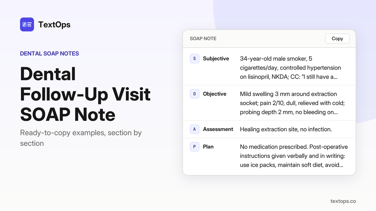
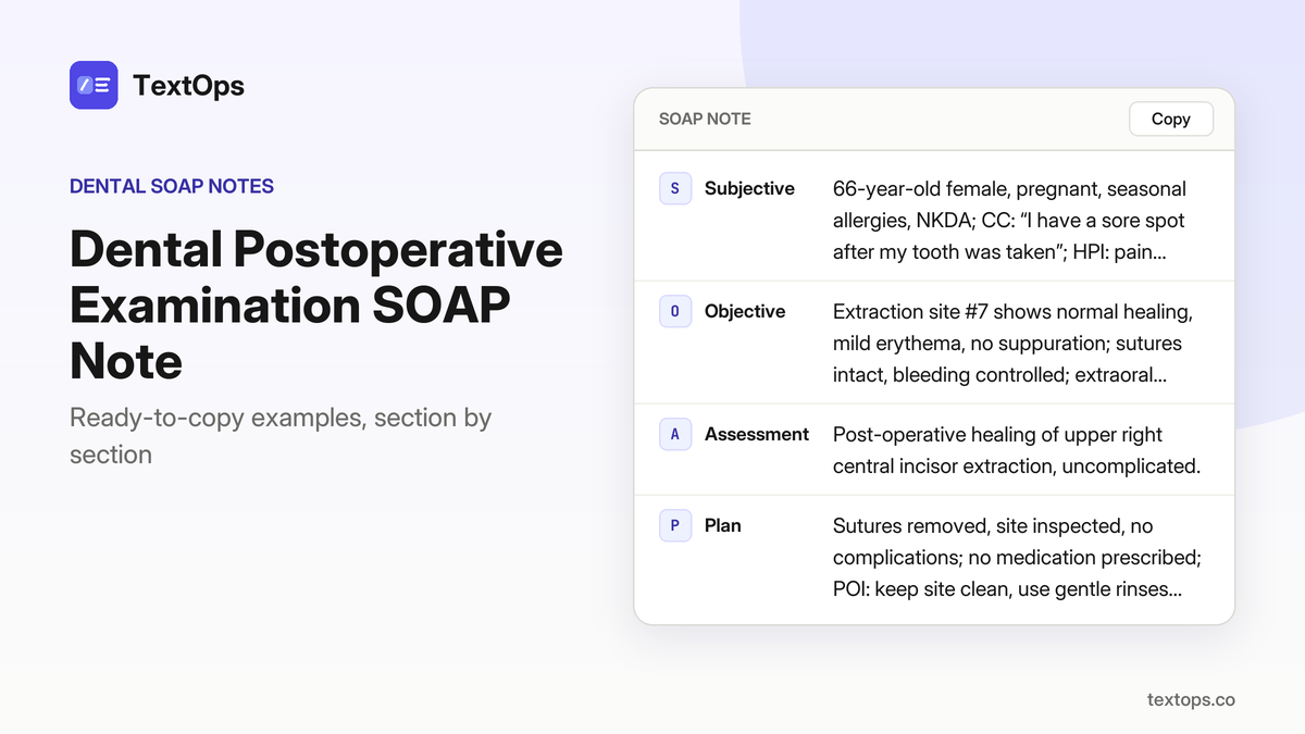
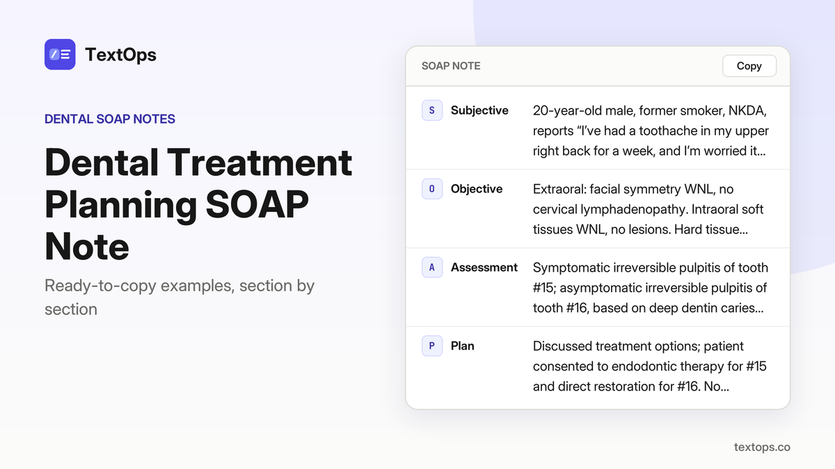
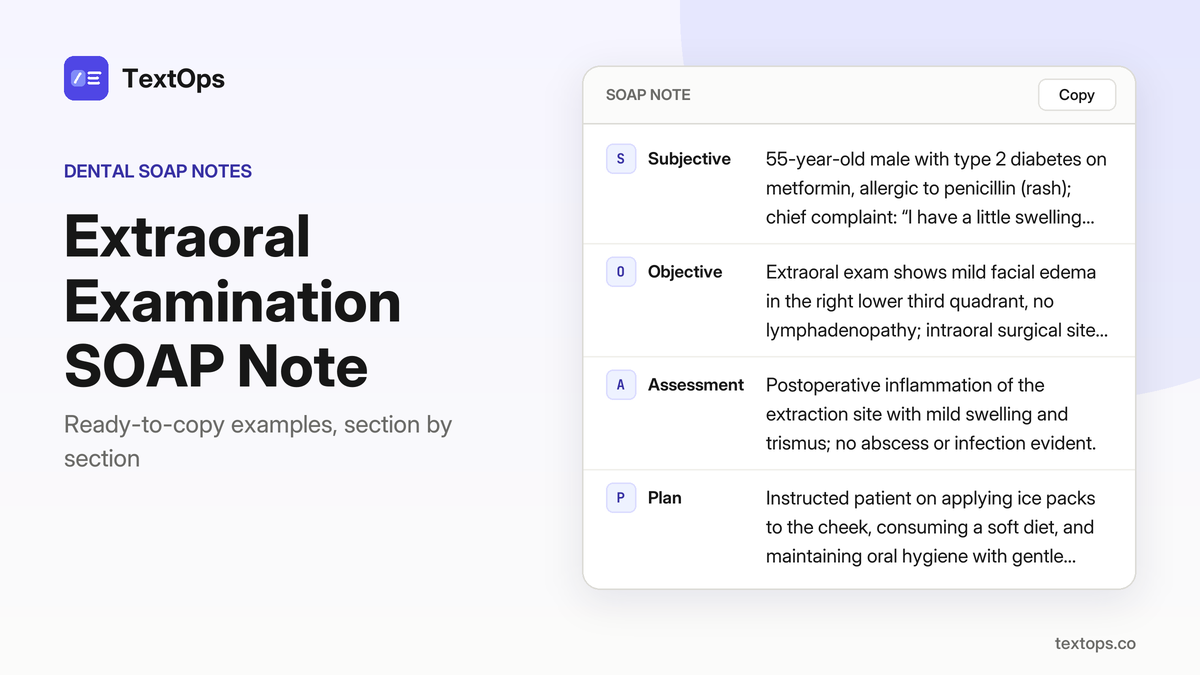
Dental radiographic SOAP notes are available for instant copy. Each note shows a full, fully filled SOAP structure, with findings placed in the Assessment section. The examples cover routine, limited, and special-population exams—ranging from routine panoramic to limited periapical for pain. Adapt the ready-to-use notes to any patient scenario.
Post-Extraction Follow-Ups
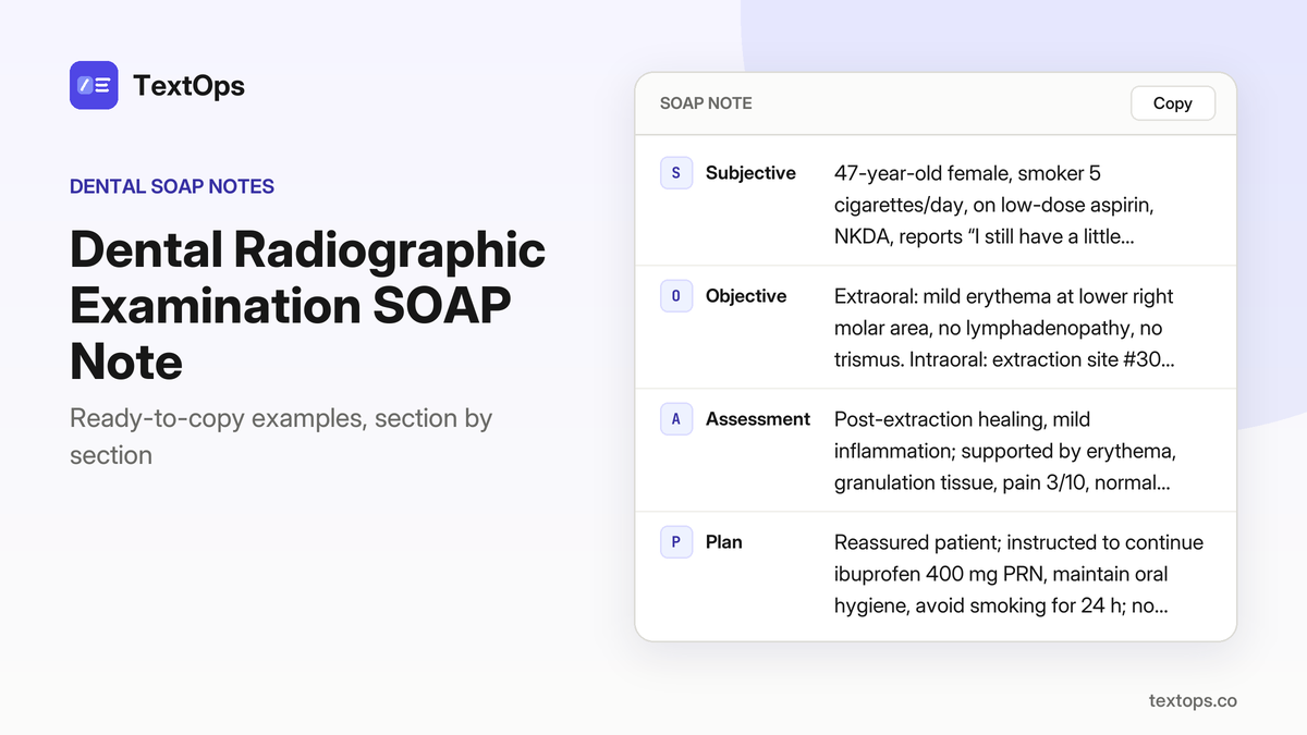
Smoker, Day 3 After Lower Molar Extraction
SOAP note
SSubjective
47-year-old female, smoker 5 cigarettes/day, on low-dose aspirin, NKDA, reports “I still have a little swelling and pain around the extraction site.” Pain 3/10, dull, relieved by ibuprofen; no systemic signs.
OObjective
Extraoral: mild erythema at lower right molar area, no lymphadenopathy, no trismus. Intraoral: extraction site #30 shows mild erythema, healing granulation tissue, no drainage. Periodontal: probing of adjacent teeth normal. Radiographic: none taken. Tests: temperature 98.6 °F.
AAssessment
Post-extraction healing, mild inflammation; supported by erythema, granulation tissue, pain 3/10, normal periodontal status.
PPlan
Reassured patient; instructed to continue ibuprofen 400 mg PRN, maintain oral hygiene, avoid smoking for 24 h; no drugs prescribed; next visit in 7 days for healing assessment.
Smoker, 50, Re-evaluation of Periapical Radiolucency
SOAP note
SSubjective
50-year-old male, smoker, asthma on albuterol PRN, NKDA; CC: “The X-ray shows a dark spot near my lower right first molar, is that a problem?”; HPI: patient reports no pain, 0/10, no swelling, no sinus tract, temperature 98.4 °F; PMH: smoker 5 cigarettes/day, asthma, no other meds.
OObjective
Periapical radiograph of tooth #30 shows a 3 mm radiolucent area at the apex; periodontal probing depth 2 mm, no bleeding on probing; extraoral exam WNL; no systemic signs noted.
AAssessment
Chronic apical periodontitis, asymptomatic, supported by 3 mm periapical radiolucency, normal probing depth, absence of pain or swelling.
PPlan
Plan root-canal therapy for tooth #30; schedule appointment in 2 weeks; no anesthesia or medications prescribed; patient advised to maintain oral hygiene and report any new symptoms immediately.
Diabetic, HbA1c 9.1, Post-Extraction Healing
SOAP note
SSubjective
37-year-old male, type 2 diabetes on metformin, smoker 5 cigarettes/day, NKDA, chief complaint: “My extraction site still feels sore, and I’m worried about healing.” HPI: pain 4/10, sharp, worsens with chewing, no trismus, no lymphadenopathy.
OObjective
Extraction socket #16 shows healthy granulation tissue, no infection; probing of adjacent teeth normal; temperature 98.7°F; HbA1c 9.1%; no systemic signs.
AAssessment
Delayed healing secondary to uncontrolled diabetes, supported by persistent soreness, sharp pain on chewing, and high HbA1c.
PPlan
Advised strict glycemic control, continue ibuprofen 400 mg PRN, avoid smoking, schedule follow-up in 7 days. No anesthesia or drugs administered today.
Smoker, 40, Emergency Re-evaluation of Tooth Fracture
SOAP note
SSubjective
40-year-old male smoker, 5 cigarettes/day, hypothyroidism on levothyroxine; CC: “I have a cracked tooth that’s hurting and I’m worried it might break off.” HPI: pain 7/10, sharp, aggravated by biting, no swelling or sinus tract, no systemic signs.
OObjective
Tooth #24 shows a vertical fracture from occlusal surface to pulp chamber; probing depth 1 mm, no BOP; temperature 98.5 °F; pain 7/10, sharp, biting aggravated; no swelling, no sinus tract; systemic signs none.
AAssessment
Symptomatic irreversible pulpitis with pulpal exposure due to fracture, supported by vertical fracture extending to pulp chamber and pain on biting.
PPlan
Plan pulpotomy with calcium hydroxide and stainless steel crown placement, schedule appointment today; no drugs prescribed; instructions to avoid hard foods, maintain oral hygiene, report any swelling or increased pain; next visit for crown placement.
Diabetic, HbA1c 7.0, Limited Bite-Wing for Post-Extraction Caries
SOAP note
SSubjective
25-year-old male, type 2 diabetes on metformin, controlled hypertension on lisinopril, NKDA, reports “I had a filling removed and I’m worried about a new cavity forming.” HPI: No pain, 0/10, no swelling, temperature 98.4 °F, HbA1c 7.0 %.
OObjective
Bite-wing radiograph of tooth #12 shows a 2 mm proximal caries extending into outer dentin; probing depth 2 mm, no bleeding on probing; no systemic signs.
AAssessment
Caries, proximal, outer dentin, supported by radiographic lesion and clinical probing.
PPlan
Recommend resin composite restoration of tooth #12; schedule restorative appointment in 1 week; no anesthesia or medication prescribed; patient instructed on oral hygiene and to report any pain or swelling immediately.
Elderly, Osteoporosis, Limited Panoramic for Osteonecrosis Screening
SOAP note
SSubjective
30-year-old female patient, seasonal allergies, NKDA; chief complaint: “I’m here for a quick check after my extraction, can you look for bone loss?”; no pain, pain score 0/10; temperature 98.6 °F; no systemic signs.
OObjective
Limited panoramic radiograph shows no osteonecrosis, bone density normal, no radiolucencies; extraoral exam: no swelling; intraoral: probing depths within normal limits; no tenderness; vital signs stable.
AAssessment
Normal post-extraction bone healing, supported by panoramic findings of intact bone density and absence of radiolucencies.
PPlan
Reassured patient; no medications prescribed; advised routine follow-up in 4 weeks; instructed to maintain oral hygiene and report any new pain or swelling.
Smoker, 45, Re-evaluation of Periodontal Disease
SOAP note
SSubjective
45-year-old male, smokes 5 cigarettes/day, allergic to penicillin (rash); chief complaint: “My gums still bleed when I brush.”; medical history: smoker; medications: none; allergies: penicillin rash.
OObjective
Periodontal probing of tooth #20 shows PD 4 mm, BOP present, CAL 3 mm; no swelling, no pain; temperature 98.5 °F; pain 1/10, mild discomfort when brushing; no systemic signs.
AAssessment
Stage II periodontitis, Grade B, supported by PD 4 mm, BOP, CAL 3 mm.
PPlan
Scaling and root planing performed; chlorhexidine gluconate 0.12% rinse 15 mL for 30 seconds, twice daily prescribed; next recall in 3 months.
Smoker, 60, Periodontal Recall with Full-mouth Series
SOAP note
SSubjective
60-year-old female, former smoker, NKDA, says “I’m here for my routine cleaning and a quick check of my gums.”
OObjective
Full-mouth series shows no new pathology, mild generalized bone loss; probing depths max 3 mm, BOP 20%; no swelling, no pain; temperature 98.4 °F; pain 0/10; no systemic signs.
AAssessment
Stage I periodontitis, Grade A, evidenced by mild bone loss, shallow probing depths, and BOP 20 %.
PPlan
Scaling and root planing performed; oral hygiene reinforcement given; next recall scheduled in 6 months.
Smoker, 55, Limited Radiographic Exam for Painful Tooth
SOAP note
SSubjective
55-year-old female, smoker, type 2 diabetic on metformin, NKDA; chief complaint: “I have a sharp pain in my lower right canine that won’t go away.” Pain 8/10, sharp, worsens with biting; no systemic signs, temperature 98.6 °F.
OObjective
Limited periapical radiograph of tooth #27 shows a 1.5 mm radiolucency at the apex; buccal mucosa adjacent to #27 is swollen; no sinus tract; probing depth 2 mm, no BOP.
AAssessment
Symptomatic apical periodontitis of tooth #27, supported by the radiolucency, pain pattern, and localized swelling.
PPlan
Plan root-canal therapy for tooth #27; prescribe ibuprofen 400 mg PRN for pain; patient advised to avoid hard foods, maintain oral hygiene, and return for RCT appointment.
Elderly, 75, Re-evaluation of Implant Site
SOAP note
SSubjective
75-year-old female, NKDA, presents with the chief complaint “I’m here to check on the implant I had placed last month,” reporting no pain or discomfort since the procedure.
OObjective
The implant site at tooth #26 shows stable bone architecture and slight pink mucosa; probing depth is 2 mm with no bleeding on probing. There is no swelling, no erythema, and no tenderness to palpation. Vital signs are within normal limits: temperature 98.5 °F, pain 0/10, and no systemic signs such as fever or malaise.
AAssessment
Successful implant integration with no evidence of peri-implant disease.
PPlan
Reassured the patient that the implant site is healthy; no local anesthesia or medication was required. Scheduled the next maintenance visit in 6 months and advised routine oral hygiene and periodic monitoring.
SOAP note
SSubjective
47-year-old female, smoker 5 cigarettes/day, takes low-dose aspirin daily, NKDA; chief complaint: “Here for my cleaning”; no pain or swelling reported; medical history: chronic smoker, aspirin use; no other systemic conditions noted.
OObjective
Extraoral examination: facial symmetry WNL, no cervical lymphadenopathy, TMJ sounds absent, maximal opening 35 mm; intraoral: periodontal probing WNL, no suppuration, no exposed bone; radiographs not taken today; all tests negative.
AAssessment
Periodic oral evaluation with no pathology identified; assessment: routine maintenance cleaning indicated.
PPlan
Full mouth prophylaxis performed; patient instructed on flossing, brushing technique, and advised to return in 6 months for next cleaning; no medications prescribed.
Routine Panoramic for Implant Screening
Elderly, 70, Routine Panoramic for Dental Implant Screening
SOAP note
SSubjective
70-year-old male patient, daily low-dose aspirin, NKDA, presents for a routine panoramic to determine implant candidacy; patient states, “Here for my routine panoramic to see if I can get an implant.”
OObjective
Panoramic radiograph shows adequate bone height and width in the posterior mandible; mild generalized bone loss consistent with age; no obvious pathology, cyst, or tumor; systemic signs are none; anesthesia not required.
AAssessment
Adequate bone for implant placement, supported by panoramic findings of sufficient height and width and absence of pathology.
PPlan
Discussed implant candidacy; planned CBCT for precise bone volume assessment; scheduled surgical consultation; no drugs administered.
Elderly, 68, Routine Panoramic for Implant Planning
SOAP note
SSubjective
68-year-old male, asthma, NKDA, states “I want to get a full implant plan, just a quick panoramic.”
OObjective
Panoramic radiograph demonstrates sufficient bone height and width in the maxillary left first molar region, with mild periodontal bone loss noted; there is no periapical radiolucency or other radiographic pathology.
AAssessment
Adequate bone for implant placement, as evidenced by the panoramic bone volume and lack of destructive lesions.
PPlan
No local anesthesia was administered. The patient was advised that a CBCT scan is required for precise bone mapping and implant site evaluation. Implant options were discussed, and a surgical consult was scheduled for definitive planning. No medications were prescribed.
Elderly, Osteoporosis, Routine Panoramic for Implant Screening
SOAP note
SSubjective
67-year-old male, smoker 5 cigarettes/day, NKDA, reports “I need a panoramic to see if I can get an implant before I quit smoking.”
OObjective
Panoramic shows moderate bone loss in the posterior mandible, mild periodontal bone loss, no cystic lesions or obvious pathology.
AAssessment
Adequate bone for implant placement with caution due to smoking, supported by panoramic findings of moderate posterior mandibular bone loss and mild periodontal loss.
PPlan
No anesthesia used. Advised smoking cessation, scheduled CBCT for precise bone assessment, discussed implant timing and potential bone grafting; no drugs prescribed.
Elderly, Osteoporosis, CBCT for Implant Planning
SOAP note
SSubjective
82-year-old male, hypothyroidism on levothyroxine, allergic to penicillin (rash); CC: “I want a panoramic to check bone before getting an implant”; PMH: hypothyroidism; no other meds; no systemic signs; no anesthesia needed.
OObjective
PAN shows reduced bone height in the maxillary right first molar region, mild generalized bone loss, no obvious pathology, bone density appears low; extraoral and intraoral exam WNL; no swelling or pain; no percussion tenderness; no periodontal probing abnormalities noted.
AAssessment
Insufficient bone height for implant placement without grafting, supported by panoramic evidence of reduced maxillary bone height and low bone density.
PPlan
Recommend CBCT for volumetric assessment of the maxillary bone; discuss bone grafting options; refer to oral surgeon for surgical planning; advise patient to continue levothyroxine; no drugs prescribed; next visit to review CBCT results and surgical plan.
Pregnancy-Related Radiographs
Pregnant, First Trimester, Routine Panoramic
SOAP note
SSubjective
38-year-old female, pregnant in the first trimester, controlled hypertension on lisinopril, NKDA, states “I just want to make sure everything looks normal.”
OObjective
Panoramic radiograph taken with a low-dose protocol shows no radiolucencies, adequate bone height, no root resorption, and no cystic lesions; extraoral examination reveals no facial asymmetry or lymphadenopathy, intraoral mucosa is intact, and periodontal probing shows no bleeding or deep pockets; no local anesthesia was required.
AAssessment
Routine panoramic, no pathology; findings confirm a normal radiographic assessment and no treatment is indicated.
PPlan
No treatment performed; patient was counseled on radiation safety during pregnancy, advised to avoid unnecessary imaging, and scheduled for a routine follow-up in 6 months; no medications prescribed.
Pregnancy, First Trimester, CBCT for Orthodontic Assessment
SOAP note
SSubjective
45-year-old pregnant female, seasonal allergies, NKDA, reports “I need to know if my tooth is impacted before I start braces.”
OObjective
CBCT shows impacted molar #15; bone density normal, no cystic changes, adequate bone volume for orthodontic movement; systemic signs none; anesthesia not used.
AAssessment
Impacted molar #15, no pathology, confirmed by CBCT findings of normal bone and absence of cystic lesions.
PPlan
Refer to orthodontist for treatment planning; no immediate intervention or drugs; counsel patient on radiation safety, noting CBCT dose is within acceptable limits for pregnancy, and schedule next visit to discuss orthodontic options.
Pregnancy, First Trimester, Routine Bite-Wing
SOAP note
SSubjective
79-year-old pregnant patient, first trimester, allergic to penicillin (rash), reports pain when biting down on tooth #12.
OObjective
Bite-wing radiograph shows proximal caries on the mesial surface of #12 extending into outer dentin; no restoration, no periapical involvement; patient is afebrile with no systemic signs.
AAssessment
Caries, class II, outer dentin, confirmed by radiographic findings.
PPlan
Applied fluoride varnish to #12; no anesthesia or medication given; patient advised to avoid excessive biting, maintain oral hygiene, and return in 6 months for reevaluation; radiation exposure was minimized by using bite-wing and shielding per pregnancy safety guidelines.
Pregnancy, First Trimester, Limited Periapical for Pulpitis
SOAP note
SSubjective
49-year-old female, pregnant, former smoker, NKDA; CC: “My lower molar hurts when I chew.” HPI: pain localized to tooth #30, triggered by chewing, no swelling, fever, or trismus, and no history of trauma.
OObjective
Periapical radiograph of tooth #30 shows no radiolucency, slight widening of the periodontal ligament space, and no root resorption. Extraoral examination is unremarkable; intraoral mucosa is healthy, and there is no facial swelling or lymphadenopathy.
AAssessment
Symptomatic irreversible pulpitis with pulp necrosis—diagnosed from the pain pattern and the PDL widening seen on the periapical film.
PPlan
Local anesthesia with 2% lidocaine with 1:100,000 epinephrine, 1.8 mL (1 cartridge), IANB; pulpotomy performed on #30. Post-operative instructions were given verbally and in written form, emphasizing oral hygiene, avoiding hard foods, and monitoring for signs of infection. No medications were prescribed. Follow-up scheduled in 2 weeks for clinical reassessment and to evaluate healing.
Pregnancy, Second Trimester, Re-evaluation of Periapical Lesion
SOAP note
SSubjective
64-year-old pregnant female, type 2 diabetes on metformin, NKDA; CC: “I have a sore spot on my lower molar.” HPI: pain localized to #18, no swelling, no systemic symptoms.
OObjective
Periapical radiograph shows a 4 mm radiolucency at the apex of #18, mild bone loss, no swelling; extraoral and intraoral exams WNL; anesthesia: 2% lidocaine with 1:100,000 epinephrine, 1.8 mL (1 cartridge), IANB.
AAssessment
Chronic apical periodontitis, previously treated, supported by the 4 mm radiolucency and mild bone loss.
PPlan
Root canal retreatment scheduled; postoperative instructions given; no drugs prescribed; next visit for retreatment completion.
Pregnancy, Second Trimester, Re-evaluation of Root Canal Treatment
SOAP note
SSubjective
66-year-old pregnant female, NKDA, reports “My upper molar still feels tender after the last visit.” History: root canal treatment on tooth #16 one month ago; no systemic symptoms.
OObjective
Periapical radiograph of #16 shows slight periapical radiolucency, mild PDL widening, no abscess or swelling; extraoral and intraoral exams normal.
AAssessment
Symptomatic apical periodontitis, previously treated, supported by radiographic radiolucency and PDL widening.
PPlan
Re-instrumentation and obturation performed on tooth #16; no local anesthesia used. Post-operative instructions given verbally; patient advised to avoid hard foods, maintain oral hygiene, report any increased pain or swelling. Next visit scheduled for 4-week follow-up to assess healing.
Pregnancy, Second Trimester, Follow-up of Endodontic Treatment
SOAP note
SSubjective
36-year-old female patient, pregnant in second trimester, reports “I feel a little discomfort when I bite.” Medical history: daily low-dose aspirin, NKDA.
OObjective
Periapical radiograph of tooth #11 shows root canal filled, no radiolucency; slight periodontal pocket 4 mm; no swelling; extraoral and intraoral examination otherwise normal.
AAssessment
Successful root canal treatment with mild periodontal pocket 4 mm, no periapical pathology.
PPlan
No anesthesia or drugs administered. Maintain oral hygiene with brushing, flossing, and a fluoride mouth rinse as instructed. Re-evaluate in 6 months.
Pregnancy, Third Trimester, Limited Radiograph for Swelling
SOAP note
SSubjective
73-year-old female patient, pregnant, asthma on albuterol PRN, NKDA, reports “I have a small swelling on my lower jaw.”
OObjective
Limited periapical radiograph of tooth #30 shows no radiolucency, normal bone density, no abscess; the swelling is confined to soft tissue only, with no signs of bone involvement or infection.
AAssessment
Soft tissue swelling, no bone involvement, confirmed by normal periapical findings and absence of radiolucency.
PPlan
Prescribed chlorhexidine gluconate 0.12% rinse, 15 mL for 30 s, twice daily; no anesthesia used. Instructed patient on rinse technique, advised to maintain oral hygiene, and to re-evaluate in 1 week for resolution of swelling.
Pregnancy, Third Trimester, Re-evaluation of Root Canal Treatment
SOAP note
SSubjective
54-year-old female patient, pregnant, smoker, NKDA, reports “My molar still hurts after treatment.” She notes persistent pain in tooth #19 since the initial root canal.
OObjective
Periapical radiograph shows a 3 mm radiolucency at the apex of #19, mild bone loss, no swelling; extraoral and intraoral exams are otherwise unremarkable.
AAssessment
Chronic apical periodontitis, previously treated, is confirmed by the 3 mm periapical radiolucency and bone loss.
PPlan
Root canal retreatment scheduled; no anesthesia or medications administered today. Post-operative instructions given verbally, including oral hygiene reinforcement and a follow-up visit in 2 weeks.
Pregnancy, Third Trimester, Follow-up of Endodontic Treatment
SOAP note
SSubjective
56-year-old female patient, pregnant, hypothyroidism on levothyroxine; chief complaint: “I still feel a slight ache when I bite.”
OObjective
Periapical radiograph of tooth #27 shows the root canal filled with no radiolucency; periodontal probing reveals a 3 mm pocket; no swelling, extraoral findings, or signs of infection; anesthesia was not administered.
AAssessment
Successful root canal treatment with mild periodontal pocket.
PPlan
Maintain oral hygiene instructions; no medication prescribed; re-evaluate in 6 months.
Diabetic Dental Visits
Diabetic, HbA1c 8.2, Periodontal Recall
SOAP note
SSubjective
44-year-old male, type 2 diabetes on metformin, controlled hypertension on lisinopril, NKDA, reports “Here for my routine cleaning and to check my gums.”
OObjective
Probing depths 3–4 mm on #20 mesial and distal with bleeding on probing; CAL 2 mm on #20; radiograph shows no bone loss beyond normal for age; no caries or restorations on #20; extraoral and intraoral otherwise within normal limits.
AAssessment
Stage II periodontitis, Grade B, supported by probing depths, BOP, and CAL on #20.
PPlan
Full-mouth scaling and root planing performed; oral hygiene instruction reinforced; no medications prescribed; next visit scheduled in 6 months for maintenance.
Diabetic, HbA1c 6.5, Routine Bite-Wing
SOAP note
SSubjective
35-year-old male with type 2 diabetes on metformin, seasonal allergies, NKDA; chief complaint: “I have a toothache when I bite down”; no systemic signs, no swelling, no pain beyond biting.
OObjective
Bite-wing radiograph shows proximal caries on #12 mesial surface extending into outer dentin; no pulpal exposure or periapical radiolucency; probing depth 1 mm, no BOP; extraoral and intraoral findings normal.
AAssessment
Caries, class II, outer dentin on tooth #12, supported by radiographic decay into outer dentin and absence of pulpal or periapical involvement.
PPlan
Restorative composite on #12 mesial surface placed today; patient instructed to avoid hard foods until completion; no medication prescribed; next visit scheduled in 4 weeks for restoration placement.
Diabetic, HbA1c 7.8, Bite-Wing for Caries Detection
SOAP note
SSubjective
48-year-old male, type 2 diabetes on metformin, allergic to penicillin, reports a dull ache in the lower right back tooth; no systemic symptoms, no swelling.
OObjective
Bite-wing radiograph shows proximal caries on the distal surface of tooth #30 extending into outer dentin; no pulpal exposure or periapical radiolucency; probing depth 2 mm, no bleeding on probing; extraoral and intraoral findings within normal limits.
AAssessment
Caries, class II, outer dentin on tooth #30, supported by radiographic evidence of proximal decay and shallow probing depth.
PPlan
No anesthesia used; patient informed that antibiotics are unnecessary; plan for composite restoration on tooth #30 distal surface; patient to return in 3 weeks for restoration.
Diabetic, HbA1c 7.2, CBCT for Implant Planning
SOAP note
SSubjective
39-year-old male, type 2 diabetes on metformin, former smoker, NKDA, states “I want to know if I can get a dental implant in the lower right quadrant.”
OObjective
CBCT of tooth #31 shows 12 mm bone height, 7 mm width, moderate density, no sinus involvement or pathology; periodontal pockets ≤4 mm, no periapical radiolucency.
AAssessment
Adequate bone for implant placement at tooth #31, as evidenced by CBCT dimensions and absence of pathology.
PPlan
Discussed implant placement plan; patient consented; referred to implant surgeon; next visit in 2 weeks for surgical appointment.
Allergic to Penicillin Cases
Allergic to Penicillin, Emergency Abscess
SOAP note
SSubjective
70-year-old male, type 2 diabetes on metformin, allergic to penicillin (rash); CC: “I have a severe pain in my lower right back tooth and a swelling that’s getting bigger”; HPI: swelling increasing, low-grade fever 99.4 °F, malaise; PMH: diabetes.
OObjective
Extraoral swelling 2 cm, no lymphadenopathy; intraoral purulent drainage from #30, buccal mucosa erythematous; PA shows well-defined radiolucency at apex of #30, cortical bone intact; probing depth 6 mm, mobility Miller 1; temperature 99.4 °F; anesthesia: 2% lidocaine with 1:100,000 epinephrine, 1.8 mL, IANB, aspiration negative.
AAssessment
Acute apical abscess of tooth #30 with pulpal necrosis, supported by purulent drainage, radiolucency, 6 mm PD, and low-grade fever.
PPlan
Incision and drainage of abscess, irrigation with saline; prescribed clindamycin 300 mg PO q8h for 7 days, acetaminophen 500 mg PO q6h PRN for 3 days; referral to endodontist for root canal therapy; POI given; next visit: endodontic treatment review.
Allergic to Penicillin, Emergency Abscess with CBCT
SOAP note
SSubjective
43-year-old male, no significant medical history, allergic to penicillin (rash); chief complaint: “My lower right molar is throbbing and I can’t keep my mouth closed”; history: acute pain, inability to close mouth, no systemic signs, temperature 98.7 °F.
OObjective
Extraoral: facial swelling 1.5 cm, no lymphadenopathy; Intraoral: purulent drainage from tooth #30, buccal mucosa erythematous; CBCT: periapical radiolucency extending 4 mm beyond cortical bone, no perforation; probing depth at #30: 5 mm; mobility: Miller grade 1; anesthesia: 2% lidocaine with 1:100,000 epinephrine, 1.8 mL (1 cartridge), IANB, aspiration negative.
AAssessment
Acute apical abscess of tooth #30 with pulpal necrosis, supported by purulent drainage, radiolucency beyond cortex, 5 mm probing depth, and mobility.
PPlan
Incision and drainage performed; irrigation done; clindamycin 300 mg PO q8h for 7 days prescribed; acetaminophen 500 mg PO q6h PRN for 3 days; referral to endodontist; postoperative instructions given; next visit: endodontic treatment review.
Allergic to Penicillin, Re-evaluation of Periapical Abscess
SOAP note
SSubjective
55-year-old male, takes low-dose aspirin, allergic to penicillin (rash); chief complaint: “I still have a dull ache in my lower right back tooth and a small swelling”; history: persistent dull ache, small swelling, no systemic symptoms; medical history: aspirin; allergies: penicillin rash.
OObjective
Extraoral: mild swelling 0.8 cm, no lymphadenopathy; Intraoral: drainage from tooth #30, buccal mucosa erythematous; Periapical radiograph: persistent radiolucency at apex of #30, slight cortical expansion; Temperature 98.6 °F; Probing depth at #30: 5 mm; Mobility: Miller grade 1; no anesthesia used.
AAssessment
Chronic apical periodontitis of tooth #30 with pulpal necrosis, supported by persistent radiolucency, drainage, mild swelling, 5 mm probing depth, and grade 1 mobility.
PPlan
Scheduled root canal therapy for tooth #30; no antibiotics prescribed; postoperative instructions given verbally and in writing; next visit: root canal treatment completion.
Allergic to Penicillin, Routine Bite-Wing
SOAP note
SSubjective
29-year-old female, asthma with albuterol PRN, allergic to penicillin (rash), presents for routine cleaning. Chief complaint: “Here for my routine cleaning.” History shows no pain, no systemic symptoms, and no recent medication changes.
OObjective
Bite-wing radiograph reveals proximal caries on the mesial surface of tooth #12, limited to outer dentin. Intraoral exam shows no restorations, healthy gingiva, probing depth 1 mm, and Miller grade 0 mobility. No extraoral findings of concern.
AAssessment
Class II caries, outer dentin on tooth #12, supported by radiographic and clinical findings.
PPlan
Applied fluoride varnish to tooth #12, provided detailed oral hygiene instructions, and advised a follow-up visit in six months to reassess caries progression. No restorative treatment indicated at this time.
Allergic to Penicillin, Emergency Re-evaluation of Fractured Molar
SOAP note
SSubjective
31-year-old male, smoker, allergic to penicillin (rash), reports “My lower right molar is cracked and I can’t bite down”; no systemic symptoms.
OObjective
Extraoral: no swelling; Intraoral: fractured crown of tooth #30, buccal mucosa intact, no pulpal exposure; Probing depth 2 mm at #30, Miller grade 0 mobility; Periapical radiograph: no periapical pathology; Anesthesia: 2% lidocaine with 1:100,000 epinephrine, 1.8 mL (1 cartridge), IANB, aspiration negative.
AAssessment
Fractured tooth #30, no pulpal involvement.
PPlan
Dental bonding of fractured surface performed; patient instructed to avoid hard foods, POI given. No medications prescribed. Next visit: follow-up in 2 weeks to assess bonding integrity.
Allergic to Penicillin, Routine Panoramic
SOAP note
SSubjective
42-year-old male with hypothyroidism on levothyroxine, reports “Just a routine check-up.”
OObjective
Panoramic radiograph demonstrates no pathology, bone density within normal limits, and no impacted teeth; intraoral examination reveals healthy gingiva, no caries, probing depths of 1–2 mm all around, and Miller grade 0 mobility.
AAssessment
No radiographic pathology; periodontal status is healthy with probing depths within normal limits.
PPlan
Performed routine periodontal maintenance, reinforced oral hygiene instructions, and scheduled a follow-up in 6 months; no medication prescribed.
Allergic to Latex Cases
Allergic to Latex, Emergency Tooth Fracture
SOAP note
SSubjective
32-year-old male, controlled hypertension, latex allergy, reports sharp pain when biting on the right back tooth. He says the pain is severe and occurs only on occlusion. No fever or swelling beyond mild buccal mucosa. NKDA aside from latex.
OObjective
Extraoral normal, temperature 98.6 °F. Intraoral: tooth #3 has a vertical fracture to the pulp chamber with exposed pulp tissue; mild buccal mucosal swelling; no sinus tract. Radiograph not taken; diagnosis based on clinical exam. Anesthesia: 2% lidocaine with 1:100,000 epinephrine, 1.8 mL buccal infiltration, aspiration negative.
AAssessment
Symptomatic irreversible pulpitis with pulpal exposure due to fracture of tooth #3.
PPlan
Extracted tooth #3 under buccal infiltration, sutured buccal mucosa, gave ibuprofen 400 mg q6h PRN for 3 days, verbal and written POI, advised avoid hard foods, return if swelling or pain worsens. Latex allergy noted for future procedures.
Allergic to Latex, Routine Bite-Wing
SOAP note
SSubjective
76-year-old female with seasonal allergies, allergic to latex, CC: “I’m here for my routine check-up and would like to see if there’s any new decay.”
OObjective
bite-wing radiograph reveals a proximal carious lesion on the mesial surface of tooth #12 extending into outer dentin; no periapical radiolucency noted; periodontal probing depths are 2 mm on all surfaces; no other carious lesions detected on remaining teeth.
AAssessment
class II caries, outer dentin on tooth #12, confirmed by radiographic evidence of a mesial proximal decay that has not yet reached the pulp.
PPlan
fluoride varnish applied to #12 under local conditions; no anesthesia required; patient advised on oral hygiene and diet modifications; recall scheduled in 6 months for caries monitoring and to reassess fluoride needs.
Allergic to Latex, Routine Panoramic
SOAP note
SSubjective
19-year-old female patient, no significant medical history, allergic to latex, reports “I’m here for my annual panoramic to check my bone levels.”
OObjective
panoramic radiograph shows adequate alveolar bone height and density around all teeth; no periapical radiolucencies; no impacted teeth; no signs of osteonecrosis; no pathology detected; soft tissue margins appear intact; no evidence of cystic or neoplastic lesions; bone loss within normal limits; overall image quality satisfactory.
AAssessment
normal panoramic radiograph, supported by bone height, density, and absence of radiolucencies or pathology; no periapical lesions, cysts, or tumors identified; bone levels within expected range for age.
PPlan
no treatment indicated; patient advised to maintain routine oral hygiene and report any new symptoms; latex allergy noted for future procedural planning; schedule next panoramic in 12 months; patient reminded to avoid latex-containing materials during future visits.
Child and Adolescent Dental Care
Child, 8 Years, Fluoride Varnish and Bite-Wing
SOAP note
SSubjective
65-year-old male, former smoker, NKDA, presents for routine check-up and caries screening; patient states, “I’m here for my regular check-up and to see if I have any new cavities.”
OObjective
Extraoral: facial symmetry WNL, no lymphadenopathy; intraoral: gingival health within normal limits, no lesions; periodontal: no BOP, PD ≤3 mm; radiographic: bite-wing shows proximal caries, outer dentin, mesial surface of tooth #14, no other pathology; tests: none performed.
AAssessment
Diagnosis: Caries, class II, outer dentin on tooth #14, supported by proximal caries on bite-wing and clinical proximity to adjacent tooth.
PPlan
Fluoride varnish applied to tooth #14; no anesthesia used; patient instructed on brushing twice daily, flossing, and return for next routine check-up in 12 months.
Child, 6, Bite-Wing for Caries Screening
SOAP note
SSubjective
6-year-old female, type 2 diabetes on metformin, NKDA; mother reports no symptoms, routine check-up.
OObjective
Bite-wing radiograph shows proximal caries, outer dentin, distal surface of tooth #12; no other caries or defects noted; gingival health within normal limits.
AAssessment
Caries, class II, outer dentin on tooth #12, supported by radiographic finding of proximal caries.
PPlan
Fluoride varnish applied to tooth #12; oral hygiene education reinforced; no medications prescribed; next visit in 6 months for routine check-up.
Child, 9, Re-evaluation of Fluoride Varnish
SOAP note
SSubjective
9-year-old male, no significant medical history, NKDA, reports “I’m back for my follow-up after the last fluoride varnish.”
OObjective
Extraoral exam shows no swelling or lymphadenopathy. Intraoral surfaces are smooth, with no carious lesions; enamel appears intact and free of decay. Gingival tissues are pink, firm, and exhibit no erythema, edema, or bleeding on probing. The previous fluoride varnish appears well-maintained, with no visible residue or discoloration. No new pathology is identified on visual or tactile examination.
AAssessment
No active pathology or diagnosis; the patient remains in good oral health status.
PPlan
Reinforced the fluoride varnish schedule by applying a new varnish application to all primary and permanent teeth, emphasizing the importance of regular preventive care. Instructed the patient and parent to maintain twice-daily fluoride toothpaste use and to avoid sugary snacks between meals. Advised a return visit in 6 months for the next varnish application and to report any new symptoms immediately. No medications were prescribed.
Structure of a Dental Radiographic SOAP Note
Subjective
Record the patient’s chief complaint in their own words, followed by a concise history of present illness that includes onset, character, duration, aggravating and relieving factors, and numeric pain rating. Include the updated past medical history, current medications, and any allergies or tobacco/alcohol use. The subjective section should capture all information that the patient reports, not the clinician’s observations.
Objective
Document vital signs when relevant, extraoral and intraoral findings with specific tooth numbers and surfaces, and the type of radiograph taken. Note the reason for the radiograph and the exact areas imaged (e.g., “PA of tooth #30, MO surface”). Do not record radiographic findings here; keep them for the Assessment. Include any additional diagnostic tests performed.
Assessment
Interpret the radiographic images and combine them with the clinical findings to state the diagnosis or clinical impression. For example, “symptomatic irreversible pulpitis of tooth #30 with periapical radiolucency.” Radiographic findings belong exclusively in this section; the Objective should only describe the imaging performed, not the results.
Plan
Outline the treatment performed, postoperative instructions, prescriptions, referrals, and the date and purpose of the next visit. Specify any patient education given and document consent or refusal when applicable. Keep the plan focused on addressing the Assessment and on clear, actionable steps for follow-up.
How Findings Influence the Plan
The Plan must answer the Assessment diagnosis directly. Include definitive treatment (root canal, extraction, monitoring), postoperative instructions, prescriptions, referrals, and the next visit date and purpose. For a symptomatic irreversible pulpitis with periapical radiolucency, a typical Plan sentence reads: “Perform endodontic therapy on tooth #30, prescribe ibuprofen 400 mg PRN, advise soft diet and chlorhexidine rinse, schedule a post-treatment recall in 7 days to assess healing.” All elements should be concise, actionable, and linked to the diagnostic impression.
Adapting the Note for Different Scenarios
Routine Radiographic Exam
Include a full-mouth series and bite-wing set. Note the patient’s age, PMH, and any medication that may affect bone metabolism. Record the radiograph type, the reason (“screening for caries and periodontal disease”), and the exact areas imaged. Keep findings in Assessment.
Limited Radiographic Exam
Focus on the complaint area. Capture a periapical or bite-wing limited to the tooth(s) in question. Specify the tooth number and surface (e.g., “PA of tooth #30, MO surface”). Document why the limited study was chosen and omit unrelated fields from the Objective.
Follow-Up or Repeat Exam
Document healing status or progression. Note the interval since the prior exam, any changes in symptoms, and the specific radiograph taken (e.g., “PA of tooth #30, follow-up after root canal”). In Assessment, compare current findings to the previous note; in Plan, state monitoring or next intervention.
Special-Population Considerations
For patients with allergies, pregnancy, or other sensitivities, record the precaution in the Subjective or Objective. Note the radiograph type chosen for safety (e.g., “PAN with lead apron for pregnant patient”) and any shielding or dose-reduction measures. Keep the structure unchanged but adapt content to reflect risk mitigation.
Frequently Asked Questions
Why are radiographic findings placed in the Assessment?
Radiographic findings are interpretive data that contribute to the clinical impression. The Assessment is the section where diagnoses and impressions are stated, so findings from PA, BWX, PAN or CBCT are summarized here as part of the diagnosis. Keeping them in Assessment preserves the distinction between objective data (the fact that a radiograph was taken) and the clinician’s interpretation, which is required for clear, contemporaneous documentation and for audit trails.
Can I record radiographic findings in the Objective section?
No. The Objective section should list the imaging performed—type, area, and reason—but not the results. Radiographic findings belong exclusively in the Assessment, where they are combined with clinical findings to form a diagnosis. Recording findings in Objective would blur the objective–subjective–assessment–plan structure and create confusion during chart review.
How do I note a patient’s allergy in the Plan?
Include the allergy as a separate sub-heading or line in the Plan, e.g., “Allergy: Penicillin—avoid amoxicillin; prescribe clindamycin if needed.” This keeps the allergy record in the Plan where medication decisions are made, ensuring that any prescribed drugs are reviewed against the allergy before administration. If the allergy influences the choice of local anesthetic or other medications, note it explicitly in the Plan line that lists the drug or agent.
These examples show structure and wording only. Record what you actually found and did, and follow your practice’s documentation requirements.
Dr. Rahul Joshi is a Specialist Orthodontist with extensive clinical experience in orthodontics, dentofacial orthopaedics, and dental aesthetics. Holding a Master of Dental Surgery (MDS) in Orthodontics and Dentofacial Orthopaedics and a Bachelor of Dental Surgery (BDS) from Manipal Academy of Higher Education, he brings expertise in orthodontic treatment planning, teeth alignment, and Invisalign treatment. With professional experience across India and the Netherlands, Dr. Joshi combines clinical knowledge with practical experience in orthodontic care, dental aesthetics, and improving patients' oral health.
Master of Dental Surgery (MDS) in Orthodontics and Dentofacial Orthopaedics — Manipal Academy of Higher Education
Bachelor of Dental Surgery (BDS) — Manipal Academy of Higher Education
Stop retyping your templates
Save any template to TextOps once, then insert it anywhere on the web without retyping. Share it with your team so everyone uses the same wording.