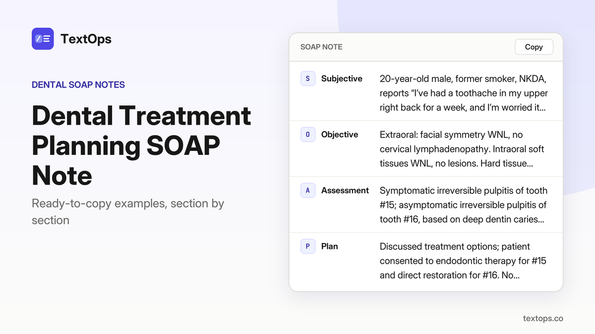
A ready-to-copy comprehensive dental exam SOAP note is presented, including subjective, objective, assessment, and plan sections. Unlike a new-patient note, this format emphasizes full periodontal, restorative, orthodontic, and radiographic documentation. The following sections cover radiographic findings, assessment categories that drive treatment planning, and template customization for varied patient scenarios.
New Patients with Medical Conditions
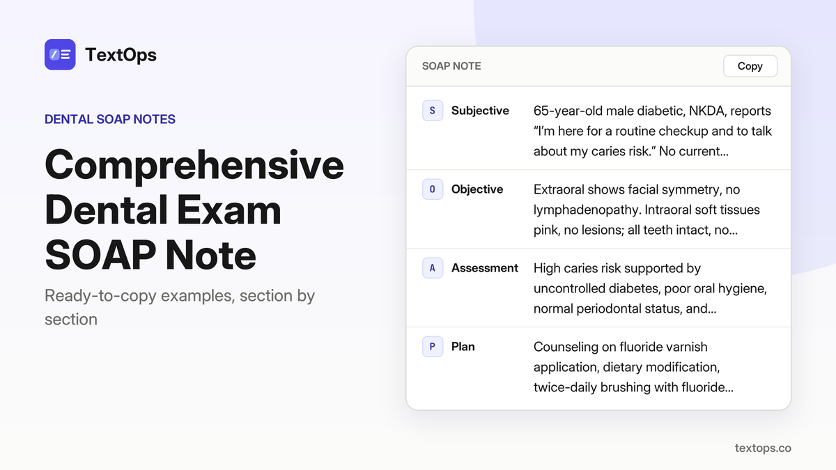
Diabetic, 65, New Patient, Caries Risk Evaluation
SOAP note
SSubjective
65-year-old male diabetic, NKDA, reports “I’m here for a routine checkup and to talk about my caries risk.” No current symptoms; medical history includes uncontrolled diabetes, no other medications.
OObjective
Extraoral shows facial symmetry, no lymphadenopathy. Intraoral soft tissues pink, no lesions; all teeth intact, no caries or restorations. Probing depths 1–2 mm, no bleeding on probing; periodontal screening score 0. Panoramic radiograph shows no periapical radiolucencies, no root resorption. Caries risk assessment high due to uncontrolled diabetes and poor oral hygiene.
AAssessment
High caries risk supported by uncontrolled diabetes, poor oral hygiene, normal periodontal status, and panoramic findings.
PPlan
Counseling on fluoride varnish application, dietary modification, twice-daily brushing with fluoride toothpaste. No anesthesia used. Patient instructed to return in 6 months for reassessment and possible fluoride varnish re-application.
Hypertensive, 60, New Patient, Orthodontic Referral
SOAP note
SSubjective
60-year-old female, hypertensive, NKDA, reports “I’d like to discuss my bite and possible orthodontic treatment.” No other medical complaints; hypertension controlled on medication.
OObjective
Extraoral: normal TMJ function, no deviation. Intraoral: Class II Division 1 malocclusion, overjet 6 mm, overbite 3 mm, upper incisors proclined, lower incisors retroclined. Probing depths 1–2 mm, no BOP; periodontal screening score 1. Panoramic radiograph: all permanent teeth present, no pathology. Cephalometric analysis shows skeletal Class II.
AAssessment
Class II Division 1 malocclusion, supported by overjet, overbite, incisor positioning and cephalometric findings.
PPlan
No local anesthesia used. Discussed orthodontic referral; explained treatment options, estimated duration, and potential need for future periodontal maintenance. Patient consented to referral. Scheduled follow-up in 4 weeks to review orthodontist’s assessment and plan.
Diabetic, 70, New Patient, Oral Cancer Screening
SOAP note
SSubjective
70-year-old male diabetic, no known drug allergies, presents for a full exam and to check for any oral cancer signs; reports “I want a full exam and to check for any oral cancer signs.” PMH includes diabetes; no other systemic issues noted.
OObjective
Extraoral exam shows no cervical lymphadenopathy. Intraoral exam reveals no lesions on lips, buccal mucosa, tongue, floor of mouth, or palate; no ulcerations or indurations. Probing depths 1–2 mm, no BOP; periodontal screening score 0. Panoramic radiograph shows no periapical radiolucencies.
AAssessment
No oral cancer detected, supported by normal soft-tissue exam and clear panoramic radiograph findings.
PPlan
No anesthesia administered. Advised routine oral cancer screening at each visit; reinforced tobacco cessation counseling. Scheduled next routine check-up in 12 months.
Elderly, 82, Post-Surgical Follow-Up
SOAP note
SSubjective
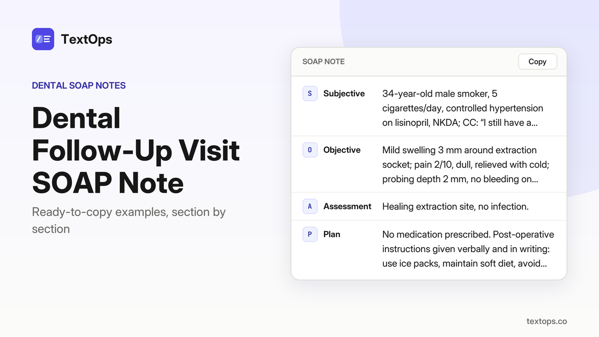
82-year-old male, asthma (albuterol PRN), NKDA, reports “I’m here for the follow-up after my tooth was removed last week.” He has no pain, mild tenderness around the extraction socket.
OObjective
Extraoral: swelling resolved, no erythema. Intraoral: extraction socket of tooth #30 healing well, no drainage, mild tenderness to palpation. Probing depths 1–2 mm around adjacent teeth; periodontal screening score 0. Panoramic radiograph: socket shows normal bone fill, no fracture.
AAssessment
Healing extraction socket, no complications, supported by absence of drainage, mild tenderness only, normal bone fill on PAN.
PPlan
No anesthesia used. Instructed on oral hygiene around socket; chlorhexidine gluconate 0.12 % rinse 15 mL twice daily for 2 weeks. Prescribed ibuprofen 400 mg every 6 hours PRN, up to 3 days for discomfort. Next visit in 2 weeks for suture removal if applicable.
Diabetic, 58, New Patient, Root Canal Therapy
SOAP note
SSubjective
58-year-old male with diabetes, NKDA, reports “my lower left first molar hurts when I bite down”; pain started 3 days ago, throbbing, worse with pressure, no swelling. PMH: type 2 diabetes, no other meds.
OObjective
Extraoral: no facial swelling. Intraoral: tooth #19 shows deep caries extending to pulp chamber, tender to percussion, no swelling; probing depth 3 mm, no BOP; periodontal screening score 1; periapical radiograph: 3 mm radiolucency indicative of symptomatic apical periodontitis; pulpal diagnosis: symptomatic irreversible pulpitis; apical diagnosis: symptomatic apical periodontitis.
AAssessment
Symptomatic irreversible pulpitis with symptomatic apical periodontitis, supported by deep caries, pulpal tenderness, 3 mm radiolucency and probing depth 3 mm.
PPlan
Administered 2% lidocaine with 1:100,000 epinephrine, 1.8 mL (1 cartridge), IANB, aspiration negative. Prepared access, removed pulpal tissue, instrumented to size 30, irrigated with 2.5% NaOCl, final rinse 17% EDTA, dried, obturated with gutta-percha and AH-plus sealer, placed temporary restoration. Prescribed ibuprofen 400 mg q6h PRN up to 3 days. Instructed on pain control, oral hygiene, avoid hard foods; scheduled post-endodontic restoration in 4 weeks.
Smoker, 60, New Patient, Oral Cancer Screening
SOAP note
SSubjective
60-year-old female smoker, NKDA, reports “I’d like a routine exam and check for any cancer signs.” She has no known medical conditions, no medications, and denies pain or other symptoms.
OObjective
Extraoral exam shows no cervical lymphadenopathy. Intraoral mucosa, lips, buccal mucosa, tongue, floor of mouth, and palate are free of lesions, ulcerations, or indurations. Probing depths are 1–2 mm with no BOP; periodontal screening score is 0. Panoramic radiograph reveals no periapical radiolucencies.
AAssessment
No oral cancer detected; findings support a normal oral cancer screening in a smoker.
PPlan
Advised routine oral cancer screening at each visit. Reinforced tobacco cessation counseling, provided written resources, and scheduled a follow-up in 6 months for re-screening. No anesthesia or medications were used.
Diabetic, 55, New Patient, CBCT for Root Canal
SOAP note
SSubjective
55-year-old male diabetic, no known drug allergies, reports “pain in my lower right first molar” and wants a detailed scan before treatment; has no other systemic symptoms.
OObjective
Extraoral exam shows no swelling; intraoral exam of tooth #30 reveals deep caries, tender to percussion, probing depth 3 mm, no bleeding on probing, periodontal screening score 1; CBCT shows a single root with two canals, slight curvature, no periapical radiolucency; pulpal diagnosis symptomatic irreversible pulpitis, apical diagnosis asymptomatic apical periodontitis.
AAssessment
Symptomatic irreversible pulpitis with asymptomatic apical periodontitis, supported by deep caries, tenderness to percussion, CBCT root morphology and absence of periapical radiolucency.
PPlan
Informed consent obtained for root-canal therapy; CBCT findings used to plan access and canal configuration; no anesthesia administered today; patient instructed on oral hygiene and pain control with NSAIDs as needed; next appointment scheduled for endodontic treatment.
Smoker, 48, New Patient, Periodontal Screening
SOAP note
SSubjective
48-year-old female smoker presents for a periodontal screening, saying “I’d like to check my gum health.” She reports no pain or bleeding and denies systemic symptoms. Past medical history is unremarkable; she takes no medications and has no known drug allergies (NKDA).
OObjective
Extraoral exam shows no lymphadenopathy. Intraoral gingiva is pink, with no recession and probing depths of 1–2 mm; no bleeding on probing. Periodontal screening score is 0 and panoramic radiograph shows no bone loss.
AAssessment
Healthy periodontium, supported by normal probing depths, absence of recession, no BOP, PSR score 0, and radiographic bone levels within normal limits.
PPlan
No local anesthesia used. Advised non-surgical periodontal maintenance every 3 months. Discussed smoking cessation and its impact on periodontal health; patient agreed to consider cessation aids. Next visit scheduled for routine maintenance.
Diabetic, 62, New Patient, Orthodontic Evaluation
SOAP note
SSubjective
62-year-old male with diabetes, NKDA, reports “I want to see if braces would help my bite.” He has no current pain, no medication changes, and denies systemic symptoms.
OObjective
Extraoral shows normal TMJ function. Intraoral reveals Class II Division 1 malocclusion, overjet 5 mm, overbite 3 mm, mild crowding; probing depths 1–2 mm, no BOP, periodontal screening score 1. Panoramic radiograph shows all permanent teeth present, no pathology. Cephalometric analysis confirms skeletal Class II.
AAssessment
Class II Division 1 malocclusion, supported by clinical overjet, overbite, crowding, and cephalometric skeletal Class II.
PPlan
Discussed orthodontic treatment options and estimated duration; no anesthesia required. Referred to orthodontist for comprehensive treatment planning. Patient advised to maintain current diabetes control and oral hygiene. Next visit: orthodontic referral follow-up.
Smoker, 54, New Patient, Implant Evaluation
SOAP note
SSubjective
54-year-old female smoker, NKDA, reports “I’d like to know if I can get a dental implant for my missing tooth.” She has no known medical conditions or medications.
OObjective
Extraoral exam shows no facial swelling. Intraoral, tooth #19 is missing; adjacent #18 and #20 are healthy. CBCT reveals 10 mm bone height and 6 mm width. Probing depths 1–2 mm, no BOP; periodontal screening score 0. Panoramic radiograph shows no pathology.
AAssessment
Adequate bone for implant placement, supported by CBCT bone dimensions and healthy surrounding periodontium.
PPlan
Discussed implant placement procedure, risks, and maintenance; no local anesthesia used today. Patient instructed on oral hygiene and scheduled for surgical appointment in 2 weeks.
Pregnancy, 29, New Patient, Dental Anxiety Management
SOAP note
SSubjective
29-year-old pregnant female presents with dental anxiety, saying “I’m nervous about dental visits during pregnancy.” She reports no pain, no prior dental issues, and no known drug allergies (NKDA). Medical history includes current pregnancy; no other systemic conditions noted.
OObjective
Extraoral exam shows normal facial symmetry. Intraoral surfaces are healthy, no caries; probing depths 1–2 mm with no bleeding on probing. Periodontal screening score is 0. Panoramic radiograph reveals no pathology.
AAssessment
Dental anxiety, supported by patient’s stated fear and absence of dental pathology or other contributory findings.
PPlan
Educated patient on safe dental care during pregnancy, emphasizing minimal radiation protocols and the safety of nitrous oxide sedation. Offered nitrous oxide for future visits; no anesthesia administered today. Advised to maintain good oral hygiene, avoid sugary foods, and attend routine check-ups. Scheduled follow-up in 4 weeks to reassess anxiety and plan any needed treatment.
Orthodontic Referral for Class II Division 1 Malocclusion in a New Patient
SOAP note
SSubjective
23-year-old female, no known drug allergies, reports “I have a noticeable overjet and would like braces.” She has no systemic symptoms, no medications, and no prior orthodontic treatment.
OObjective
Extraoral: normal TMJ function, facial symmetry, no lymphadenopathy. Intraoral: Class II Division 1 malocclusion, overjet 6 mm, overbite 3 mm, mild crowding; probing depths 1–2 mm, no BOP, periodontal screening score 1. Panoramic radiograph shows all permanent teeth present, no pathology; cephalometric analysis confirms skeletal Class II.
AAssessment
Class II Division 1 malocclusion, supported by 6 mm overjet, 3 mm overbite, mild crowding, and skeletal Class II cephalometric findings.
PPlan
Referred to orthodontist for comprehensive evaluation and treatment planning. Discussed options (fixed brackets, clear aligners) and estimated duration (≈24 months). No local anesthesia or medication administered. Advised oral hygiene instruction, routine recall in 4 weeks for orthodontic referral follow-up.
Allergic to Aspirin, 55, Emergency Abscess Drainage
SOAP note
SSubjective
55-year-old male reports “a painful swelling on my lower right molar” that started yesterday; he describes throbbing pain that worsens with chewing. PMH includes aspirin allergy (rash); no other drug allergies (NKDA).
OObjective
Extraoral exam shows diffuse 2 cm swelling, tender to palpation, no fever. Intraoral: tooth #30 has deep caries, purulent drainage, tender to percussion; probing depth 4 mm, BOP present; periodontal screening score 3. Periapical radiograph reveals a 5 mm periapical radiolucency with abscess formation; no other findings noted.
AAssessment
Pulp necrosis with acute apical abscess, supported by deep caries, purulent drainage, 4 mm PD with BOP, and a 5 mm periapical radiolucency.
PPlan
Incision and drainage of abscess performed under 2% lidocaine with 1:100,000 epinephrine, 1.8 mL (1 cartridge), IANB, aspiration negative. Socket curetted, irrigated with saline, temporary restoration placed. Prescribed clindamycin 300 mg PO q8h for 7 days (due to aspirin allergy) and ibuprofen 400 mg PO q6h PRN up to 3 days. Instructed on oral hygiene, avoid hot foods, return for root-canal therapy referral.
Smoker, 52, New Patient, Orthodontic Evaluation
SOAP note
SSubjective
52-year-old female smoker presents with “I’d like to see if braces would help my bite.” She reports no pain or trauma. Past medical history is unremarkable; she has no known drug allergies (NKDA).
OObjective
Extraoral exam shows normal TMJ function. Intraoral findings reveal a Class II Division 1 malocclusion with overjet 5 mm, overbite 3 mm, and mild crowding. Probing depths 1–2 mm, no BOP; periodontal screening score 1. Panoramic radiograph shows all permanent teeth present, no pathology. Cephalometric analysis confirms skeletal Class II.
AAssessment
Class II Division 1 malocclusion, supported by overjet, overbite, crowding, and skeletal Class II findings.
PPlan
No local anesthesia was administered. Discussed orthodontic referral, treatment options, and estimated duration. Provided written instructions on oral hygiene during treatment. Scheduled follow-up with orthodontist; patient to return to clinic for comprehensive evaluation.
Allergy-Specific Comprehensive Exams
Allergic to Penicillin, 12-Year-Old, Dental Trauma
SOAP note
SSubjective
12-year-old male, asthma (albuterol PRN), NKDA except penicillin allergy, reports “my dad said I need a checkup, but I have a sore tooth.”
OObjective
Extraoral: facial symmetry, no lymphadenopathy. Intraoral: mild gingivitis, one enamel caries on tooth #15 occlusal; periodontal PSR 0; no other caries.
AAssessment
Enamel caries on tooth #15 and mild gingivitis, supported by the occlusal caries and gingival inflammation noted on exam.
PPlan
Applied fluoride varnish to tooth #15; instructed on brushing twice daily, flossing, and avoidance of sugary snacks; no anesthesia used; scheduled follow-up in 4 weeks for caries removal plan.
Allergic to Latex, 40, Post-Extraction Review
SOAP note
SSubjective
40-year-old female, reports “I still feel a bit sore after the extraction.” She is on low-dose aspirin daily, has no known drug allergies, and is allergic to latex.
OObjective
Extraoral exam shows no lymphadenopathy. Intraoral findings: healing socket at #18, soft tissue intact, no bleeding, mild swelling. Periodontal assessment normal.
AAssessment
Healing extraction site, no infection, supported by intact soft tissue, absence of bleeding, and lack of signs of infection.
PPlan
No anesthesia used. Oral hygiene instruction given; prescribed chlorhexidine gluconate 0.12% rinse, 15 mL for 30 s twice daily for 7 days. Follow-up scheduled in 7 days.
Allergic to Penicillin, 22, Periodic Exam
SOAP note
SSubjective
43-year-old female smoker, 5 cigarettes/day, presents for routine cleaning. She reports no pain or concerns. PMH: NKDA, allergic to penicillin; no other medical issues.
OObjective
Extraoral exam shows facial symmetry, no lymphadenopathy. Intraoral surfaces exhibit mild plaque accumulation, no carious lesions, and no oral mucosal abnormalities. Periodontal screening code PSR 1 (mild gingivitis).
AAssessment
Mild gingivitis (Periodontitis Stage I, Grade A) supported by PSR 1, mild plaque, and absence of lesions or mobility.
PPlan
Scaling and polishing performed without local anesthesia. Oral hygiene instruction given; fluoride varnish applied to all surfaces. Patient advised to maintain twice-daily brushing, flossing, and return for routine exam in 6 months.
Allergic to Latex, 45, New Patient, Caries Risk
SOAP note
SSubjective
28-year-old female, “I want to see if I need fillings.” Reports no pain, has type 2 diabetes on metformin, no other meds, allergic to latex.
OObjective
Extraoral normal. Intraoral: one enamel caries on tooth #30 occlusal surface; periodontal PSR 0; caries risk high due to diabetes and poor oral hygiene; no radiographs taken.
AAssessment
Enamel caries on tooth #30, high caries risk—supported by the single enamel lesion and diabetes status.
PPlan
Applied fluoride varnish to #30; discussed caries removal plan and reinforced oral hygiene; no anesthesia used. Advised to return in 6 months for reassessment.
Allergic to Penicillin, 38, Periodic Exam, Oral Hygiene
SOAP note
SSubjective
60-year-old female, “I want to keep my teeth clean.” She reports no pain or discomfort; she has controlled hypertension on lisinopril, no other medications, and is allergic to penicillin.
OObjective
Extraoral examination shows facial symmetry, no lymphadenopathy, and normal TMJ function. Intraoral findings include moderate plaque accumulation, generalized gingival inflammation, no caries, and no oral lesions. Periodontal screening (PSR) is 1, indicating mild gingivitis.
AAssessment
Gingivitis (Periodontitis Stage I, Grade A) – supported by moderate plaque, gingival inflammation, and PSR 1.
PPlan
Scaling and polishing performed without local anesthesia. Fluoride varnish applied to all tooth surfaces. Patient received detailed oral hygiene instruction, emphasizing twice-daily brushing with a soft-bristle brush, interdental cleaning, and reduction of sugary snacks. Advised to return in 6 months for periodontal maintenance and reassessment of plaque control.
Allergic to Latex, 50, Periodic Exam, Oral Hygiene
SOAP note
SSubjective
49-year-old male presents for a periodic oral evaluation with the chief complaint “just a routine checkup.” He reports no pain, no swelling, no recent trauma, and no systemic illnesses; he is not taking any medications and has no known drug allergies except a documented latex allergy, which has caused mild dermatitis in the past and is managed by avoiding latex gloves and instruments.
OObjective
Extraoral examination reveals facial symmetry, no cervical or submandibular lymphadenopathy, and a normal range of motion of the temporomandibular joint. Intraoral inspection shows mild plaque accumulation on the buccal surfaces of the anterior teeth, no carious lesions, no active periodontal pockets, and a periodontal screening recording (PSR) of 0 indicating healthy periodontium. No oral mucosal lesions, ulcers, or erythema are noted.
AAssessment
The patient has a healthy periodontium, as evidenced by plaque control, a PSR score of 0, and the absence of gingival inflammation or periodontal pockets.
PPlan
Polishing was performed with a latex-free polishing pad and a non-latex polishing paste, followed by the application of a fluoride varnish to all tooth surfaces. Oral hygiene instruction was reinforced, emphasizing proper brushing technique, flossing, and the use of a soft-bristled brush. Latex-free instruments were used throughout to prevent allergic reaction. The patient was advised to return for a routine periodic exam in six months and to contact the office sooner if any new symptoms develop.
Allergic to Penicillin, 60, New Patient, Oral Cancer Screening
SOAP note
SSubjective
32-year-old female, chief complaint: "I want a cancer screening." No pain or symptoms reported. Past medical history: hypothyroidism on levothyroxine. Allergic to penicillin (rash); NKDA otherwise.
OObjective
Extraoral exam shows facial symmetry, no lymphadenopathy. Intraoral soft-tissue survey: lips, buccal mucosa, tongue, floor of mouth, palate, gingiva all within normal limits; no ulcerations or lesions. Periodontal probing within normal limits. No radiographs taken.
AAssessment
No oral cancer detected; findings of normal mucosa and gingiva support this.
PPlan
Routine comprehensive exam performed. Oral hygiene instruction given; fluoride varnish applied. No anesthesia used. Patient advised to return in 12 months for periodic exam.
Elderly, 75, Periodic Exam, Prosthetic Evaluation
SOAP note
SSubjective
69-year-old female, former smoker, NKDA, allergic to penicillin; CC: “My dentures feel loose.” HPI: reports gradual loosening over 3 months, no pain, no swelling; PMH: no systemic disease.
OObjective
Extraoral: facial symmetry, no lymphadenopathy; Intraoral: partial denture #19-#21 poorly fitting, palatal mucosal irritation, mild plaque; no caries; periodontal PSR 0; no radiographs taken.
AAssessment
Prosthetic failure with mucosal irritation, supported by loose denture fit and palatal tissue inflammation.
PPlan
Adjusted denture fit intraorally; reinforced oral hygiene instructions with gentle brushing; no medication prescribed; patient advised to avoid latex products during future procedures; schedule follow-up in 4 weeks.
Elderly, 80, Periodic Exam, TMJ Assessment
SOAP note
SSubjective
80-year-old female, established patient, presents for a periodic oral evaluation with chief complaint “My jaw hurts when I open my mouth.” She reports a dull, aching pain that has been present for the past two weeks, worsens with chewing, and is relieved by over-the-counter ibuprofen. She has a history of seasonal allergies, no known drug allergies, and is allergic to latex.
OObjective
Extraoral: TMJ clicking on opening, limited maximum opening 35 mm, mild pain on palpation; no swelling, no lymphadenopathy. Intraoral: no lesions, mucosa pink and intact, no caries or restorations noted. Periodontal: all teeth present, probing depths 1–2 mm, no bleeding on probing. Radiographic: periapical radiographs of the mandibular molars show no pathology.
AAssessment
TMJ disorder, Stage I, Grade A – limited opening, clicking, and pain on palpation.
PPlan
No anesthesia administered. Prescribed soft diet, jaw relaxation exercises, and referred to a physical therapist for TMJ management. Advised to avoid hard foods and to use heat packs as needed. Follow-up in 4 weeks for reassessment of pain and function.
74-year-old female reports “My denture is uncomfortable.” She has no significant medical history, NKDA, and is allergic to penicillin with a rash reaction. HPI: discomfort for 2 months, worsening when chewing, no pain.
OObjective
Extraoral: facial symmetry, no lymphadenopathy. Intraoral: partial denture with poor fit, mucosal inflammation on posterior palatal region; mild plaque, no caries; periodontal PSR 0. No radiographs taken.
AAssessment
Prosthetic maintenance required; findings of poor fit and palatal inflammation support this diagnosis.
PPlan
Adjusted denture to relieve pressure, reinforced oral hygiene with brushing and interdental cleaning instructions, and scheduled follow-up in 4 weeks. No anesthesia or medications were administered.
Elderly, 83, Periodic Exam, TMJ Management
SOAP note
SSubjective
50-year-old male, chief complaint: “My jaw hurts after chewing.” History: pain began 2 weeks ago, worsens with chewing, no prior TMJ issues. Medical history: asthma, uses albuterol PRN; no other meds. Allergies: latex, no other drug allergies.
OObjective
Extraoral: no swelling, no lymphadenopathy; TMJ: clicking, limited opening 32 mm, pain on palpation. Intraoral: no lesions; periodontal status WNL; no radiographs taken.
AAssessment
Stage I TMJ disorder, Grade A, supported by clicking, limited opening, and pain on palpation.
PPlan
Fabricated custom occlusal splint; instructed jaw relaxation exercises; referred to physical therapy for TMJ management. No anesthesia used. POI: wear splint nightly, perform exercises 3×/day; next visit in 4 weeks for splint adjustment and PT follow-up.
Smoker-Related Comprehensive Exams
Smoker, Day 3 After Lower Molar Extraction
SOAP note
SSubjective
33-year-old female, “I’m still feeling a bit sore and the swelling is a little worse than yesterday.” History: day-3 post-extraction of lower molars #29-#31, takes low-dose aspirin, NKDA.
OObjective
Extraoral: no trismus, no lymphadenopathy. Intraoral: extraction socket of #30 shows mild erythema, 2 mm soft-tissue edema, no purulent drainage. Periodontal: probing of #29 and #31 1 mm PD, no bleeding. Radiograph: bitewing shows no periapical radiolucency.
AAssessment
Healing extraction socket, no complications; supported by mild erythema, minimal edema, normal probing and clear radiograph.
PPlan
No anesthesia administered. Instructed on oral hygiene; prescribed ibuprofen 400 mg every 6 h as needed up to 3 days, chlorhexidine 0.12 % rinse 15 mL 30 s twice daily. Advised to avoid smoking until socket fully healed; next visit in 7 days for follow-up.
Elderly, 78, Periodontal Disease Progression
SOAP note
SSubjective
78-year-old male smoker reports “my gums feel more swollen and I notice more bleeding when I brush.” He has a 5-cigarette-daily habit, no known drug allergies, and no other medical conditions.
OObjective
Extraoral exam is unremarkable. Intraoral periodontium shows PSR 3 on teeth #17, #18, #19; BOP 80 %; probing depths 5–6 mm buccally; CAL 4 mm; horizontal bone loss 3 mm on bitewing radiographs; no furcation involvement, no mobility, no systemic signs of infection.
AAssessment
Periodontitis Stage III, Grade B, supported by PSR 3, 80 % BOP, 5–6 mm PD, 4 mm CAL, and 3 mm horizontal bone loss around #17–#19.
PPlan
Initiated scaling and root planing on #17–#19 (no anesthesia). Prescribed chlorhexidine 0.12 % rinse, 15 mL, 30 s, twice daily. Advised strict smoking cessation counseling, oral hygiene instruction, and scheduled reassessment in 4 weeks.
Smoker, 55, Dental Implant Evaluation
SOAP note
SSubjective
30-year-old female, smoker, reports “I’d like to know if I can get an implant in place of my missing upper left canine.” She has type 2 diabetes, well controlled (HbA1c 6.5 %), takes metformin, NKDA.
OObjective
Extraoral normal. Intraoral: missing tooth #11, surrounding bone appears adequate. Periodontal: no inflammation, probing depths ≤3 mm. Panoramic radiograph shows 12 mm bone height from crest to sinus floor and 6 mm width. CBCT indicates bone density 1200 HU, no sinus pathology. No signs of infection.
AAssessment
Adequate bone for implant placement in the upper left canine area, supported by 12 mm height, 6 mm width, 1200 HU density, and absence of sinus or infectious pathology.
PPlan
Discussed implant placement plan, obtained informed consent, scheduled surgical placement in 2 weeks. No anesthesia given today. Post-operative instructions: maintain oral hygiene, avoid smoking for 48 h, return for surgical placement.
Smoker, 50, Periodic Exam with New Caries
SOAP note
SSubjective
50-year-old male smoker reports “I have a new spot on my front tooth that’s getting darker.” He has controlled hypertension on lisinopril, NKDA, and no other medical issues.
OObjective
Extraoral exam normal. Intraoral: dark brown lesion on facial surface of tooth #7, 3 mm diameter, involving enamel and outer dentin; no sensitivity to percussion. Periodontal probing WNL. Bitewing radiograph shows no radiolucency or proximal caries.
AAssessment
Caries, enamel and outer dentin, Black’s class II of tooth #7, confirmed by clinical appearance and radiograph.
PPlan
Applied resin composite restoration to tooth #7 without anesthesia. Instructed patient on fluoride toothpaste, interdental cleaning, and smoking cessation counseling; scheduled next visit in 6 months for maintenance.
Smoker, 48, Periodic Exam, Plaque Index
SOAP note
SSubjective
55-year-old female, NKDA, presents for routine cleaning, stating “I just came in for my routine cleaning.” No significant medical history, no medications, no allergies.
OObjective
Extraoral findings normal. Intraoral plaque index 3.5/5 overall, highest on buccal surfaces of lower molars. Probing depths 1–2 mm all around, no BOP. Bitewing radiographs show no new caries. No periodontal disease noted.
AAssessment
Plaque accumulation with mild gingivitis, evidenced by high plaque index and normal probing depths without BOP.
PPlan
Performed scaling and polishing. Instructed on brushing technique and advised chlorhexidine 0.12% rinse, 15 mL, 30 s, twice daily. No anesthesia used. Next visit scheduled in 6 months for routine recall.
Root Canal Therapy for a Severely Carious Premolar with Trismus
SOAP note
SSubjective
59-year-old male reports “upper left premolars hurt a lot and I can’t open my mouth wide.” He has hypothyroidism on levothyroxine, is allergic to penicillin (rash), NKDA otherwise.
OObjective
Extraoral: facial symmetry, no lymphadenopathy. Intraoral: tooth #17 shows deep caries extending to outer dentin with pulpal exposure, spontaneous pain, percussion pain; tooth #18 has deep caries, pulpal exposure, pain on biting. Probing depth 1–2 mm, no BOP. Radiograph: radiolucency at apices of #17 and #18 indicating chronic apical periodontitis. Trismus: maximal opening 30 mm. No systemic signs of infection.
AAssessment
Symptomatic irreversible pulpitis with symptomatic apical periodontitis of teeth #17 and #18, supported by pulpal exposure, spontaneous and percussion pain, and periapical radiolucency.
PPlan
Administered 2% lidocaine with 1:100,000 epinephrine, 1.8 mL (1 cartridge) IANB. Obtained pulpal access, performed root canal therapy on #17 and #18, placed calcium hydroxide intracanal medication, obturated with gutta-percha and sealer, restored #17 with composite, placed temporary on #18. Prescribed ibuprofen 400 mg PRN. Instructed on pain control, oral hygiene, and to return in 7 days for final restoration.
Pregnancy-Related Comprehensive Exams
Pregnant, First Trimester, Routine Check-Up
SOAP note
SSubjective
28-year-old pregnant patient, first trimester, presents for routine check-up; states “Here for my routine check-up.” Medical history: none; NKDA. No medications or allergies noted. HPI is uncomplicated and limited to a routine review.
OObjective
Extraoral examination shows facial symmetry, no lymphadenopathy, and normal TMJ function. Intraoral assessment reveals excellent oral hygiene, no plaque, no caries, and no restorations. Periodontal probing depths are 1-2 mm bilaterally with no bleeding on probing. Panoramic radiograph taken with minimal exposure shows no pathology, and all soft tissues appear normal.
AAssessment
Routine comprehensive exam; the absence of caries, restorations, periodontal disease, and radiographic pathology confirms a low caries risk status.
PPlan
Fluoride varnish applied to all surfaces; oral hygiene instructions reinforced with emphasis on diet and plaque control; no anesthesia required. The patient was reassured about radiation safety in early pregnancy and advised to avoid unnecessary imaging. Next visit scheduled in 6 months for routine follow-up.
Pregnancy, 32, Emergency Pain Management
SOAP note
SSubjective
32-year-old pregnant female reports “My lower molar hurts badly, I think it’s a toothache.” She describes throbbing pain radiating to the right ear, lasting >24 h, with swelling. PMH: none, NKDA, no medications.
OObjective
Extraoral: facial symmetry normal, no lymphadenopathy. Intraoral: swelling in right mandibular molar region, percussion tender over tooth #30. Periodontal: no bleeding on probing, probing depths within normal limits. Radiograph: periapical PA shows radiolucency at apex of #30. Temperature 99.5 °F.
AAssessment
Pulp necrosis with acute apical abscess of tooth #30, supported by throbbing pain, swelling, percussion tenderness, radiolucency at apex, and low fever.
PPlan
Administered 2% lidocaine with 1:100,000 epinephrine, 1.8 mL (1 cartridge), IANB; aspiration negative. Drained abscess, irrigated site. Prescribed amoxicillin 500 mg q8h for 7 days, ibuprofen 400 mg q6h PRN for 5 days. Informed patient of antibiotic indications, oral hygiene, and signs of worsening infection. Scheduled follow-up in 48 h for reassessment.
Pregnant, 30, Routine Check-Up, CBCT Indicated
SOAP note
SSubjective
30-year-old female, pregnancy status confirmed, reports “I want a CBCT for my wisdom teeth.” Past medical history non-contributory, NKDA. HPI: routine check-up, no pain or swelling.
OObjective
Extraoral: facial symmetry, no lymphadenopathy. Intraoral: soft tissues intact, no lesions. Periodontal: no probing depths >4 mm, no BOP. Radiographic: periapical film shows impacted tooth #30 with root resorption; no other pathology. No tests performed.
AAssessment
Impacted third molar with root resorption, confirmed by periapical radiograph and indicated for CBCT evaluation of root morphology and bone volume.
PPlan
Discussed CBCT benefits and radiation safety in pregnancy; obtained informed consent. No anesthesia used. Scheduled CBCT appointment; advised to avoid additional x-rays until CBCT is completed. Next visit: review CBCT results and plan extraction if indicated.
Pregnancy, 35, Dental Implant Planning
SOAP note
SSubjective
63-year-old female, asthma controlled with as needed albuterol, NKDA. Chief complaint: “I want an implant in the upper right canine.” History: missing tooth #6, adjacent teeth intact, no pain or swelling. Medical history: asthma, no other chronic conditions.
OObjective
Extraoral: facial symmetry, no lymphadenopathy. Intraoral: tooth #6 absent, adjacent teeth intact, no caries or periodontal pockets. Periodontal: no attachment loss, BOP 0 %. Radiographic: CBCT shows bone height 10 mm, width 8 mm, adequate density; panoramic radiograph shows no pathology. Tests: none performed.
AAssessment
Edentulous site with adequate bone for implant placement, confirmed by CBCT bone dimensions and absence of periodontal disease.
PPlan
Discussed implant placement options and timing; no local anesthesia required today. Advised oral hygiene instructions, no medication prescribed. Scheduled surgical appointment for implant placement; patient to return for pre-op clearance.
44-year-old female, chief complaint: “I’m on aspirin, can I get a cleaning?” History: takes low-dose aspirin daily, no known drug allergies, no other systemic issues.
OObjective
Extraoral: facial symmetry, no lymphadenopathy. Intraoral: mucosa pink, no lesions. Periodontal: probing depths 1-2 mm, no bleeding on probing, no calculus or plaque accumulation. Radiographs: none taken.
AAssessment
Routine exam with anticoagulant review; findings of healthy gingiva and no calculus support continuation of aspirin therapy.
PPlan
No anesthesia used. Advised continuation of aspirin, use of rubber dam for prophylaxis, application of fluoride varnish. Patient instructed on oral hygiene, advised to avoid extractions or procedures that increase bleeding risk. Next visit scheduled in 6 months for routine check-up.
Pregnant, 32, Periodic Exam, Oral Cancer Screening
SOAP note
SSubjective
46-year-old male, presents for a periodic exam, “Just a routine check, no pain.” He reports smoking about five cigarettes a day, has no known drug allergies, and denies any systemic symptoms or recent illnesses. Medical history is otherwise unremarkable.
OObjective
Extraoral exam shows facial symmetry, no cervical or submandibular lymphadenopathy, and normal TMJ function. Intraoral mucosa is pink, smooth, and free of ulcerations or lesions; lips, buccal mucosa, tongue, floor of mouth, hard and soft palate, and oropharynx are all within normal limits. Periodontal probing reveals depths of 1–2 mm bilaterally with no bleeding on probing. There are no carious lesions, no restorations, and no missing teeth. No radiographs were taken during this visit.
AAssessment
Routine exam with oral cancer screening; the normal mucosal appearance, absence of lesions, and healthy periodontal status support a negative oral cancer screen.
PPlan
No local anesthesia was administered. Discussed the risks of continued smoking, reinforced cessation strategies, and provided written educational materials on oral health and smoking. Advised the patient to return for a routine check-up in six months to continue monitoring periodontal health and oral cancer screening.
Diabetic-Related Comprehensive Exams
Diabetic, 68, Periodic Exam, Periodontal Therapy
SOAP note
SSubjective
77-year-old male, type 2 diabetes on metformin, NKDA, reports “here for my routine cleaning and to check my gums.” He has no pain or swelling, and his diabetes has been stable with recent HbA1c <7%.
OObjective
Extraoral: facial symmetry, no lymphadenopathy. Intraoral: healthy mucosa, no lesions. Periodontal: probing depths 3 mm on teeth #15-#17, BOP present on #15 and #16, CAL 2 mm on #15, plaque index 2, no mobility, no furcation involvement. Radiographic: panoramic shows mild generalized bone loss consistent with stage I periodontitis; no periapical pathology.
AAssessment
Periodontitis Stage I, Grade A, supported by PD 3 mm, BOP on #15-#16, CAL 2 mm, and radiographic bone loss <1 mm.
PPlan
Scaling and root planing performed on #15-#17; no anesthesia used. Prescribed chlorhexidine gluconate 0.12% rinse, 15 mL, 30 seconds, twice daily for 7 days. Advised strict oral hygiene instruction and to maintain HbA1c <7%. Next periodontal maintenance scheduled in 3 months.
54-year-old male, “Just a routine check-up, no pain.” PMH: controlled hypertension on lisinopril; NKDA.
OObjective
Extraoral: facial symmetry, no lymphadenopathy. Intraoral: plaque index 3, no caries, no mobility, no furcation involvement. Periodontal: PD 4 mm on #11-#13, BOP on #12, CAL 3 mm on #11. Radiographs: PA shows slight periapical radiolucency on #12 (chronic apical periodontitis); PAN shows generalized bone loss stage II periodontitis.
AAssessment
Periodontitis Stage II, Grade A, and chronic apical periodontitis on tooth #12, supported by PD/BOP/CAL values and radiographic findings.
PPlan
Scaling and root planing performed on #11-#13; chlorhexidine 0.12% rinse 15 mL, 30 seconds, twice daily for 7 days prescribed. No anesthesia used. Follow-up periodontal reassessment in 4 weeks; discussed hypertension control and maintaining BP <140/90.
Non-Surgical Periodontal Therapy for Recurrent Gingival Recession
SOAP note
SSubjective
71-year-old male, “My gums keep receding around my front teeth,” reports progressive recession over several months; no pain, no systemic disease, NKDA, no medications.
OObjective
Extraoral: facial symmetry normal, no lymphadenopathy. Intraoral: recession 4 mm on facial surface of tooth #7, probing depth 3 mm, BOP present, CAL 2 mm, no mobility, no furcation involvement. Radiographs: periapical PA shows no pathology; panoramic PAN shows adequate bone height. Plaque index 2, no caries, no restorations on #7.
AAssessment
Recurrent gingival recession on tooth #7 with Periodontitis Stage I, Grade A, supported by 4 mm recession, 3 mm PD, BOP and 2 mm CAL.
PPlan
Performed scaling and root planing on tooth #7; prescribed chlorhexidine gluconate 0.12 % 15 mL rinse, 30 s, twice daily for 7 days; instructed gentle brushing technique to avoid aggressive brushing; scheduled periodontal maintenance in 3 months; discussed smoking cessation and its impact on recession.
Documenting Radiographic Findings in a Comprehensive Exam
Periapical (PA) radiographs are taken when a specific tooth or group of teeth is suspected of pulpal or periapical pathology; note the tooth number(s) and the finding, e.g., “PA of tooth #15 shows a 3 mm radiolucent periapical lesion.” Bitewing (BWX) films are indicated for caries detection in the posterior dentition; record the quadrants and surfaces, e.g., “BWX of upper right quadrant shows proximal caries on #16/17, MO surface.” A full-mouth series (FMX) is used when a comprehensive assessment of all teeth is required, such as before restorative work or orthodontic planning; document the series and any notable findings, e.g., “FMX shows missing tooth #30, restoration on #19, and mild attrition on #25.” Panoramic (PAN) radiographs are indicated for evaluating the entire arch, impacted teeth, or jaw pathology; record the indication and key observations, e.g., “PAN reveals an impacted canine in the left maxilla and a 2 mm radiolucency in the right mandibular body.” Cone-beam CT (CBCT) is reserved for complex cases requiring three-dimensional detail, such as implant placement or root canal anatomy; note the scan area and findings, e.g., “CBCT of the maxillary left molar region shows a Type II root canal configuration and a 1 mm cortical bone defect.”
PA – single tooth or small group; note tooth number(s) and lesion size or density.
BWX – posterior surfaces; list quadrants and surfaces with caries or restoration status.
FMX – all teeth; record series and any gross findings (missing, restoration, wear).
PAN – entire arch; indicate reason (impaction, jaw pathology) and key observations.
CBCT – 3-D detail; specify scan area, anatomical variants, bone defects, or pathology.
If the patient declines a radiograph, document the refusal and explain diagnostic limits.
Assessment Categories and Their Role in Treatment Planning
Periodontal assessment records PD, CAL, BOP, and mobility for all teeth. Findings such as 4 mm PD with 2 mm CAL and BOP on tooth #15 indicate Stage II, Grade B periodontitis. Treatment plan: non-surgical scaling and root planing, chlorhexidine rinses twice daily, recall in 3 months for re-probe.
Restorative assessment charts caries by tooth, surface, and depth. A 3 mm enamel caries on the buccal surface of tooth #29 requires a silver-ion fluoride sealant. Plan: apply sealant, reinforce oral hygiene education, schedule next visit in 12 months.
Orthodontic assessment evaluates malocclusion, overjet, overbite, crossbite, and tooth alignment. A 5 mm overjet with a Class II canine relationship on the right side suggests orthodontic referral. Plan: refer to orthodontist, discuss treatment options, obtain consent, and schedule referral appointment.
Endodontic assessment identifies pulpal and periapical pathology. A 2 mm radiolucent periapical lesion on tooth #16 with symptomatic irreversible pulpitis requires root canal therapy. Plan: perform RCT with 2 % lidocaine + 1:100,000 epinephrine, place calcium hydroxide dressing, schedule obturation in 1 week.
Oral cancer screening inspects lips, buccal mucosa, tongue, floor of mouth, and palate. A 6 mm ulcerated lesion on the lateral border of the tongue warrants biopsy. Plan: refer to oral medicine, obtain biopsy, discuss follow-up.
Radiographic assessment selects PA, BWX, FMX, PAN, or CBCT based on findings. A missing tooth #30 and a proximal caries on tooth #27 seen on FMX informs restorative planning. Plan: document FMX findings, schedule restorative treatment for tooth #27, consider implant for tooth #30 after periodontal evaluation.
Category
Typical Findings
Example Plan
Periodontal
4 mm PD, 2 mm CAL, BOP
Scaling, chlorhexidine, 3-month recall
Restorative
3 mm enamel caries, buccal #29
Sealant, 12-month follow-up
Orthodontic
5 mm overjet, Class II canine
Orthodontic referral, consent
Endodontic
2 mm periapical lesion, symptomatic pulp
RCT, calcium hydroxide, obturation
Oral Cancer
6 mm ulcerated tongue lesion
Biopsy, oral medicine referral
Radiographic
Missing #30, proximal caries #27
FMX documentation, restorative plan
Customizing the Comprehensive SOAP Template for Different Patient Scenarios
Add a PMH line that lists chronic diseases, current medications, and anticoagulant use.
Include a vital sign check when indicated (BP for hypertension, capillary glucose for diabetes).
Document ASA status if the patient is medically complex.
In the O section, note any systemic signs that may influence dental treatment (e.g., edema, pallor, or cyanosis).
Assessment should reflect how the medical condition affects oral health (e.g., “diabetes → increased periodontal risk”).
Plan must address medical-related precautions: schedule appointments in the morning, avoid anticholinergic drugs, and coordinate with the physician for medication adjustments.
Vital signs, extraoral findings, intraoral soft tissue, periodontal probing (PD, CAL, BOP), caries chart, radiographic findings (PA, BWX, FMX, PAN, CBCT), oral cancer screening, tobacco use (cigarettes/day), gestational age
AAssessment
Diagnosis or clinical impression incorporating medical condition (e.g., Stage II periodontitis in diabetic patient, smoker with oral cancer risk, pregnant patient with bleeding gums)
PPlan
Treatment plan addressing dental and medical needs (e.g., periodontal therapy, smoking cessation counseling, safe medication list, timing of elective procedures, coordination with physician)
Allergy-Specific Comprehensive Exams
Add the allergy in the S line and avoid known allergens in the O and P sections.SOAP note
A new-patient SOAP note is a first-visit snapshot; it captures the full medical and dental history, vital signs, and a baseline oral examination, but it does not include a detailed periodontal chart or a full restorative assessment. The comprehensive exam expands the objective section to full periodontal probing, complete caries charting, occlusal analysis, and a full-mouth radiographic series, and it adds a treatment-planning phase that integrates medical risk factors into a long-term care plan. Typical findings in a new-patient note are a general health summary, chief complaint, and initial visual screening, whereas a comprehensive note documents probing depths, CAL, BOP, caries locations, and radiographic findings that guide definitive therapy.
Unique elements in a new-patient note
Full medical history and medication list
Vital signs (BP, pulse) when indicated
Baseline oral cancer screening
Initial caries risk assessment
General periodontal screening (PSR or probing if warranted)
Baseline radiographs (e.g., bitewing or panoramic)
Treatment plan focused on preventive and initial care steps
Patient education and consent documentation for future procedures
These examples show structure and wording only. Record what you actually found and did, and follow your practice’s documentation requirements.
Dr. Rahul Joshi is a Specialist Orthodontist with extensive clinical experience in orthodontics, dentofacial orthopaedics, and dental aesthetics. Holding a Master of Dental Surgery (MDS) in Orthodontics and Dentofacial Orthopaedics and a Bachelor of Dental Surgery (BDS) from Manipal Academy of Higher Education, he brings expertise in orthodontic treatment planning, teeth alignment, and Invisalign treatment. With professional experience across India and the Netherlands, Dr. Joshi combines clinical knowledge with practical experience in orthodontic care, dental aesthetics, and improving patients' oral health.
Master of Dental Surgery (MDS) in Orthodontics and Dentofacial Orthopaedics — Manipal Academy of Higher Education
Bachelor of Dental Surgery (BDS) — Manipal Academy of Higher Education
Stop retyping your templates
Save any template to TextOps once, then insert it anywhere on the web without retyping. Share it with your team so everyone uses the same wording.